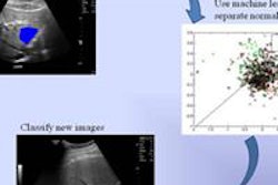
Ultrasound computer-aided detection (CAD) technology can enhance the performance of radiologists in characterizing focal liver lesions (FLLs) on contrast-enhanced ultrasound (CEUS), according to a study by Japanese researchers presented at the recent 2010 RSNA meeting.
The research team from Kumamoto University in Kumamoto found that its CAD application turned in 76.6% overall sensitivity for differentiating five types of focal liver lesions on dynamic contrast ultrasound, and it improved the performance of all seven radiologists in their study.
"Overall diagnostic accuracy of all observers was improved from 52% to 66%," said Junji Shiraishi, PhD. Shiraishi presented the research during a multisession course at the RSNA show, and co-author Katsutoshi Sugimoto, MD, PhD, also presented the findings during a separate multisession course.
Believing that CAD can compensate for the operator dependence of ultrasound, the researchers performed an observer study to demonstrate its potential in the differential diagnosis of liver lesions on contrast ultrasound.
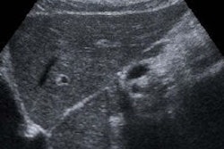
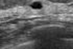
The study evaluated 107 focal liver lesions from 106 cases, including 27 metastases, 27 hemangiomas, and 53 hepatocellular carcinomas (HCCs). The 56 hepatocellular carcinomas included 17 described as well-differentiated, 26 as moderately differentiated, and 10 as poorly differentiated. Biopsy or surgical specimens were used to determine the pathology of all cases except for hemangiomas.
Dynamic contrast-enhanced images were generated using the Sonazoid contrast agent (GE Healthcare, Chalfont St. Giles, U.K.) and an Aplio XG ultrasound scanner (Toshiba Medical Systems, Otawara, Japan) with B-mode, vascular imaging with microflow imaging (less than 60 seconds after injection) and Kupffer phase (10 minutes after injection).
First without CAD, seven radiologists with experience in liver contrast ultrasound first gave their confidence ratings as to whether the lesions were benign or malignant (and if malignant, they described them as either metastasis or hepatocellular carcinoma). They also selected one of five classifications matched to the unknown focal liver lesion.
They then immediately read the cases again with the use of the institution's CAD technique for classifying focal liver lesions. The researchers analyzed the observers' rating data with multireader, multicase receiver operator characteristics (ROC) analysis.
CAD turned in an overall sensitivity of 76.6% (82/107) for differentiating the five categories of focal liver lesions in their database. In addition, all seven observers saw their mean area under the ROC curve (AUC) values for accurate characterization of benign or malignant lesions significantly increase with CAD; the mean values increased from 0.861 ± 0.068 without CAD to 0.942 ± 0.045 with CAD (p < 0.001).
The researchers also found a significant improvement in correct description of the focal liver lesions as metastasis or hepatocellular carcinoma, with mean AUC values climbing from 0.707 ± 0.09 to 0.776 ± 0.075 following use of CAD (p = 0.035). Overall diagnostic accuracy for all observers increased from 52.6% to 66.5%.
By Erik L. Ridley
AuntMinnie.com staff writer
December 17, 2010
Related Reading
Inexperienced readers benefit most from breast US CAD, March 5, 2010
Color Doppler US predicts survival of breast cancer patients, January 26, 2010
Fuzzy 3D ultrasound CAD sharpens breast cancer sensitivity, September 3, 2009
Breast US image analysis helps predict metastasis, July 29, 2009
Breast ultrasound CAD performance varies in ethnic populations, September 5, 2008
Copyright © 2010 AuntMinnie.com