CHICAGO - Researchers utilizing different imaging techniques determined that light exercise such as walking and bowling may protect people against developing osteoarthritis, while the location of fat in the body -- especially visceral or belly fat -- may contribute to the bone weakening seen in osteoarthritis.
The research was presented at RSNA annual meeting being held this week in Chicago.
Light exercise appears to improve knee cartilage as visualized in T2-weighted images from 3-tesla MRI studies; however, high-impact sports such as running and skiing are associated with knee cartilage damage in people who are overweight, said Keegan Hovis, a research associate in the department of radiology at the University of California, San Francisco (UCSF).
Hovis and colleagues enrolled 132 subjects at risk of developing knee osteoarthritis who were part of the long-term National Institutes of Health (NIH) Osteoarthritis Initiative. The group consisted of individuals who had suffered knee injuries, have a family history of osteoarthritis, or have other risk factors, according to Hovis. The researchers also enrolled 33 individuals of similar age and body habitus who were not at risk of osteoarthritis as controls. Ninety-nine women and 66 men participated, and they ranged in age from 45 to 55 years.
The subjects were stratified into three exercise levels: sedentary; light exercisers who engaged in activities such as walking or playing Frisbee for less than two hours a day, three days a week; or moderate to strenuous exercisers who engaged in running, cycling, or other sports more than an hour a day, three or more days a week. Hovis and colleagues also analyzed whether activities that require knee bending affected knee cartilage.
The T2 intensity decreased among the subjects who were at risk of osteoarthritis and engaged in light exercise, Hovis said in a press briefing. The scans also showed that women with osteoarthritis risk factors who fell into the moderate-to-strenuous exercise category had substantially more collagen degeneration in their knees than any other group.
She said that the reason for a lack of effect in men doing strenuous exercise was unknown, but it could be related to the fact that women appear more at risk of osteoarthritis than men. The researchers determined that in people who were not at risk for osteoarthritis, the level of exercise performed did not appear to affect cartilage degeneration.
However, frequent knee bending -- climbing 10 or more flights of stairs per day or kneeling for 30 or more minutes a day, for example -- was associated with more damaged cartilage in people with and without osteoarthritis risk factors.
"We're not saying you shouldn't run or climb stairs," added researcher Thomas Link, MD, chief of musculoskeletal imaging at UCSF. "But you have to be extremely careful to protect your cartilage starting at a very early age."
The No. 1 thing you can do to protect your bone health is to shed excess pounds and maintain a healthy weight, said David Levin, MD, professor and chairman emeritus of radiology at Thomas Jefferson University Hospital in Philadelphia.
That was the point made by Miriam Bredella, MD, assistant professor of radiology at Massachusetts General Hospital/Harvard Medical School in Boston. "We found that in young women, deep belly fat is bad for the bones," she said. "We now know that abdominal obesity needs to be included as a risk factor for osteoporosis and bone loss."
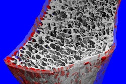
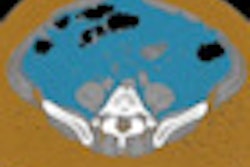
Bredella said that not all fat is the same, illustrating with MR spectroscopy and quantitative CT that women with excessive amounts of visceral fat have a more porous bone structure than women without visceral fat. On the other hand, women with excessive subcutaneous fat show normal bone structure.
She and colleagues enrolled 50 premenopausal women who were obese, meaning they had a body mass index greater than 30. They under went MR spectroscopy to evaluate bone marrow fat of the fourth vertebra in the lumbar section of the spine. The researchers used CT to determine bone mineral density in young obese women.
The researchers found that more fat present in bones correlated to lower bone mineral density. Also, the more visceral fat -- having an "applelike" figure or fat around the waist -- the more likely the woman was to have lower bone mineral density.
Bredella showed that bone mineral density was approximately 170 mg/cm3 if a woman in the study had 50 cm2 of visceral fat, but bone mineral density declined to around 140 mg/cm3 if a woman had 250 cm2 of visceral fat (p = 0.03).
"Visceral fat has damaging effects on bone health," she said in the press briefing. "Excessive deep belly fat is not only a risk factor for heart disease and diabetes but also for bone loss."
Mary Mahoney, MD, professor of radiology at the University of Cincinnati, noted that there is no proven treatment or dietary supplement that can preferentially get rid of belly fat rather than subcutaneous fat. "The best strategy is not to gain fat in the first place or to get right of as much body fat as possible," she said.
By Edward Susman
AuntMinnie.com contributing writer
November 30, 2010
Related Reading
Marathoners have altered biochemistry in knee cartilage, August 16, 2010
MRI can identify osteoarthritis risk factors 10 years early, June 30, 2010
Cartilage recovers quickly from a tough run, May 14, 2008
Omega-3 fatty acids linked to denser bones in men, March 23, 2007
Copyright © 2010 AuntMinnie.com