CHICAGO - While FDG-PET/CT may be more suitable for early response detection of bone involvement after stem cell transplantation, whole-body MRI can be an "additional predictive marker" for long-term evaluation, according to a study presented Sunday at the RSNA annual meeting.
Whole-body MRI and FDG-PET/CT both have been used to evaluate the initial extent of disease in multiple myeloma. German researchers noted, however, that there has been limited data about the role of the imaging modalities in post-treatment assessment.
The study, led by Christoph Weber, MD, from the University of Hamburg, analyzed 30 patients (11 women, 19 men) with an average age of 55.3 years. The patients were imaged with both PET and MRI (within one month of each scan) after stem cell transplantation at a median follow-up of 28 months.
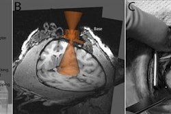
FDG-PET/CT was acquired using a standard protocol. Whole-body MRI was acquired on a 1.5-tesla system (Achieve, Philips Healthcare, Andover, MA).
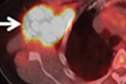
Whole-body MRI characterized bone involvement in terms of pattern, size, and contrast enhancement, while FDG-PET/CT reviewed bone involvement by pattern, size, and tracer accumulation in 10 regions of the skeleton. Extraosseous lesions also were classified.
The results showed that of the 30 patients, 22 were in complete remission, while the remaining eight patients were in partial remission. FDG-PET/CT detected a total of 10 lesions in six patients, while whole-body MRI detected a total of 97 lesions in 20 patients. In addition, 42 of the 97 contrast-enhanced lesions were detected in 11 of the 20 patients. Weber noted that the average size of the lesions was 2.2 cm.
"Overall, between FDG-PET/CT and whole-body MRI, we found concordant results in five of 102 bone lesions assignable to the multiple myeloma," Weber added.
On a per-patient basis, FDG-PET/CT had a sensitivity of 50%, compared with whole-body MRI's sensitivity of 88%. FDG-PET/CT achieved specificity of 91%, compared with 41% for whole-body MRI.
In addition, positive predictive value for FDG-PET/CT was 67%, compared with 35% for whole-body MRI. FDG-PET/CT achieved a negative predictive value of 83%, compared with 90% for whole-body MRI.
FDG-PET/CT and whole-body MRI can contribute to a more comprehensive evaluation of multiple myeloma patients after stem cell transplantation, Weber and colleagues concluded. Both modalities add information about the extent and activity of disease, with FDG-PET/CT better able to detect disease activity.
"In treated patients, the positive predictive value of MRI was disappointingly low," Weber added. "FDG-PET/CT, despite its limited capacity in detecting lesions in the post-treatment setting, might be more suitable for the response detection of bone involvement than MRI."
"Nevertheless," he continued, "showing all these lesions, whole-body MRI may be an additional predictive marker for long-term follow-up after stem cell transplantation."
By Wayne Forrest
AuntMinnie.com staff writer
November 28, 2010
Related Reading
Whole-body MRI helps predict course of asymptomatic myeloma, March 18, 2010
Contrast MRI helps guide stem cell therapy for myeloma, February 11, 2010
New progress in multiple myeloma treatment; MDCT has edge in detection, June 18, 2007
No survival benefit of high-dose therapy in multiple myeloma, January 5, 2006
Copyright © 2010 AuntMinnie.com