CHICAGO - An award-winning research project conducted by a medical student at the University of Pennsylvania Medical Center in Philadelphia found that whole-body FDG-PET added little additional information in staging patients with known primary breast cancer.
"We found that very few women are presenting with distant metastases at the time of diagnosis" said Dr. Caitlin Carr-Lopez, who discussed the results of her group's research at the RSNA conference on Sunday afternoon.
Carr-Lopez, whose work was presented with the RSNA trainee research prize - medical student, evaluated the impact of whole-body FDG-PET in staging breast cancer beyond the breast and axilla.
The scientists enrolled 200 women with breast cancer in an institutional review board-approved multimodality imaging trial conducted between March 2002 and May 2005 at their facility. The comprehensive imaging workup on the patient cohort included an initial film-screen mammogram with or without ultrasound; digital mammography, whole-breast ultrasound, MRI, and whole-body FDG-PET were then conducted on the group.
Radiologists on the team classified distant lesions on a 1 to 5 scale with 1 indicating no uptake, 2 showing physiologic uptake, 3 demonstrating nonsuspicious uptake, 4 signifying a suspicious uptake without a workup, and 5 calling a suspicious uptake with a workup. The authors wrote in their electronic poster presentation that class 5 lesions were evaluated with CT, MR, bone scan, x-ray, or pathology, and that standard staging was conducted with chest x-ray and bone scans as determined by clinical judgment.
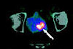
A total of 189 out of the 200 women in the study had PET scans, which were conducted on an Allegro PET system by Philips Medical Systems of Andover, MA. According to the research presented by the scientists, FDG-PET identified 71 distant areas of increased uptake in 22% of the patients. Of these 71 lesions, 59 were considered suspicious in 33 of the women.
The group classified 50 of the 59 distant lesions as class 5 and the remaining nine lesions as class 4. Out of the group of 50 class 5 lesions, 19 were found to be true-positive for neoplastic disease, two were true-positive for non-neoplastic disease, and 29 were determined to be false-positive.
The researchers reported a positive predictive value for class 5 lesions by FDG-PET of 40%. The true-positive malignancies found by FDG-PET were in six patients, or 3% of the 189 women who had a PET scan; the scientists noted that four of these patients also had their true-positive distant lesions identified by standard staging procedures, meaning that PET alone identified true-positive distant disease in only 1% of the patients.
The false-positive findings on PET in 12% of the study group resulted in 17 CTs, eight x-rays, two MRIs, and one bone scan being conducted; although one of the MRI exams revealed an additional neoplasm that had been a false negative in PET.
"In our study group, we found that there was a low positive predictive value of FDG-PET to stage breast cancer beyond the breast and axilla," Carr-Lopez said. "We also found that the high false-positive rate led to unnecessary, costly, and sometimes invasive follow-up studies."
By Jonathan S. Batchelor
AuntMinnie.com staff writer
November 26, 2006
Related Reading
Preop FDG-PET spots axillary involvement in breast cancer, may obviate biopsy, August 21, 2006
Dual-time-point breast PET increases diagnostic accuracy, December 2, 2005
PEM turns in solid results for detecting breast malignancies, November 10, 2005
PET mammography unit shows promise for DCIS, June 22, 2005
PET sharpens treatment plan for advanced breast cancer, August 16, 2004
Copyright © 2006 AuntMinnie.com