VIENNA - Will digital breast tomosynthesis (DBT) replace mammography for breast cancer screening? The answer is "maybe," and it depends on how well the modality performs in some forthcoming clinical trials, say two experts who will share their views with ECR attendees at this afternoon's special focus session.
The sensitivity of mammography for detecting breast cancer is less than optimal, primarily because the breast is a 3D structure that is projected onto a 2D radiographic image. This means normal breast tissue can conceal a tumor. DBT is a 3D radiographic technique that reduces the effect of overlapping tissues in breast cancer detection.
 The T-MIST trial should answer the question as to whether digital breast tomosynthesis can replace digital mammography, according to Dr. Martin Yaffe, from the University of Toronto in Canada.
DBT appears to be gaining in popularity. The technique improves the accuracy of finding cancers and also reduces the recall rate of women with suspicious findings, especially in women in younger women and those with dense breasts, according to Dr. Martin Yaffe, a senior scientist of imaging research at Sunnybrook Research Institute and a professor in the departments of medical biophysics and medical imaging at the University of Toronto in Canada.
"That's what the excitement is all about," he said in an interview with ECR Today.
DBT is exciting because it's the newest and most realistic competitor to digital mammography that has come up in years, according to fellow speaker Dr. Sophia Zackrisson, an associate professor of diagnostic radiology at Lund University, Skåne University Hospital in Malmö, Sweden.
"It is a similar technique to digital mammography, with images that are like digital mammography, so the radiologists can easily adapt their reading of this new technique," she said. "It is also easily integrated in the clinical setting. This is in contrast to MRI, for instance, which is more expensive, time-consuming, and more challenging in interpretation."
In tomosynthesis, the x-ray tube moves over a range of angles about a pivot point located above the digital detector to obtain a series of low-dose digital projection radiographs, Yaffe explained. The detector may be stationary or rotate about the pivot point. The x-ray tube may temporarily halt as each projection is acquired or may move continuously during acquisition.
A computer algorithm reconstructs a 3D image. The images are usually viewed as a "movie-loop" in which adjacent x-y planes are displayed sequentially. Imagers are able to see structures within the breast without overlap. The tumor is clearer, easier to see, and separate from structures in the breast.
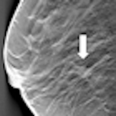
 In this screening case, an asymptomatic woman has a tumor that is not discernible on digital mammography (left), but appears as a clearly visible spiculated lesion on digital breast tomosynthesis (white arrow), which shows the slice where the lesion is in focus. The lesion is a 13-mm invasive ductal carcinoma, grade 2. Images courtesy of Dr. Sophia Zackrisson.
"Tomosynthesis from what we've seen is promising in the North American context, where a high percentage of women get called back," he said. "We can reduce that rate by 30% or more, as well as find some cancers that have not been detected."
Tomosynthesis' most likely application is in breast cancer screening, but its precise role will only be determined after a study that is comparing full-field digital mammography with DBT, the Tomosynthesis: Comparison of Full Field Digital Mammography with Digital Breast Tomosynthesis (T-MIST) trial, a multivendor, multisite trial. The trial is only for sites in North America, but will have implications for countries on other continents as well.
"Personally, I do not think it [DBT] should be used in screening before we have evidence from trials showing that we gain in detection and not lose too much on the false-positive side," Zackrisson said.
During his presentation, Yaffe will explain how tomosynthesis works, point to preliminary results of the T-MIST trial, show images, and explain why tomosynthesis is better than conventional projection. In Zackrisson's lecture, ECR delegates will get a good overview of DBT and hear results from recent studies with updates on the accuracy of the technique.
"Aspects of reading time and image presentation will be discussed, which is important since this is one of the major obstacles at least in screening," she said. "What views should be used? How should the image stacks in digital breast tomosynthesis be presented? Can we find a presentation mode which is acceptable in the screening workflow?"
DBT is a promising tool in breast imaging, she concluded. However, further evidence from the ongoing trials is needed to establish its place in breast cancer diagnosis and screening.
Originally published in ECR Today on 10 March 2013.
Copyright © 2013 European Society of Radiology