Rupture of the pectoralis major muscle is rare, but occurs notably among athletes who weightlift, either as a sport or a training regimen. And when it comes to evaluating these injuries, studies show that MR imaging is an accurate tool.
Studies of the injury have shown that a majority of incidences occur among weightlifters and bodybuilders, with most acute injuries occurring during the bench press. The injury has also been seen in contact sports, particularly football and rugby, but cuts across all athletes with recent trends toward weight training.
Pectoralis injuries are sufficiently rare that a 1999 article from New York City's Hospital for Special Surgery touted a "relatively large" sample of 15 patients. The study confirmed the suggestion of earlier reports that MR imaging of the pectoralis was highly preferable to clinical assessments, which "may be misleading and are often affected by hemorrhage, tenderness, or spasm in the acute setting," according to the study authors (Radiology, March 1999, Vol. 210:3, pp. 785-791).
A set of case studies out of the University of Iowa in 1996 focused on the utility of T1- versus T2-weighted axial images for pectoralis major rupture (Skeletal Radiology, October 1996, Vol. 25:7, pp. 625-628).
In an evaluation of the four case studies, the authors determined that diagnosis of a complete rupture was difficult in some cases with T1-weighted images because hematomas and hemorrhages were indistinguishable from the adjacent muscle. They found that T2-weighted images were effective in these cases, and that T1 images were adequate for diagnosing chronic cases.
 |
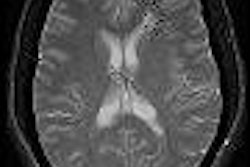
| An axial fast SE T2-weighted image [TR/effective TE (ms) 3100/91.3] shows disruption of the pectoralis major muscle near its attachment to the humerus. The linear low signal structure extending medially from the humerus was confirmed at surgery to represent torn residual tendon. |
 |
| The SE T1-weighted image [TR/TE (ms) 616/11] at the same level as figure above does not clearly show the ruptured site. This was thought to be due to the gap being filled with a hematoma having a similar signal intensity as the muscle. Images courtesy of Dr. Kenjirou Ohashi, department of radiology, University of Iowa College of Medicine; reprinted with permission from Skeletal Radiology, October 1996, Vol. 25:7, pp. 625-628. |
In the 1999 New York study, nine patients went on to primary surgical repair, and all the MR findings were confirmed at surgery, according to the study authors, led by radiologist Dr. David Connell.
"In particular, periosteal stripping was identified at the normal insertion site in all patients with complete tendon avulsion. The amount of tendon retraction correlated with the MR imaging findings," they wrote.
Technical considerations in MRI of the pectoralis include use of surface-coil and oblique-plane images obtained along the plane of the muscle-tendon unit, the authors noted.
"Recognition of abnormal signal intensity surrounding the cortex of the humerus is important because this finding is typically due to stripping of the periosteum as the tendon is avulsed from the osseous insertion," they wrote. "Owing to the proximity of the biceps tendon to the insertion of the pectoralis major muscle, one should be careful not to misinterpret abnormal signal intensity and hematoma at this location as an injury of the biceps muscle."
Getting an MR evaluation of the injury can be particularly important in determining whether surgery would be beneficial, the authors noted.
"Preoperative diagnosis of the location of the tear is important because tendon avulsion from the humerus is treated with prompt surgical repair, whereas muscle-tendon injuries are generally treated in a conservative manner," they wrote. "Early surgical repair allows avoidance of adhesions, muscle scarring, fibrosis, and atrophy, and hastens the return of the athlete to competition."
By Matt King
AuntMinnie.com contributing writer
August 25, 2004
Related Reading
MR highlights venous compression in TOS patients, August 19, 2003
Correct positioning during mammography improves breast cancer detection, March 27, 2002
Copyright © 2004 AuntMinnie.com