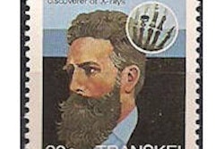
By the time news stories about Wilhelm Conrad Roentgen's discovery of the mythical "x-rays" were published in early January 1896, many physicists started using their own generators, static machines, induction coils, vacuum tubes, photographic plates, and tungsten filaments to try to produce sharper and more precise x-ray images.
A few days after Roentgen's x-ray discovery was published in a Vienna newspaper on January 11, William Randolph Hearst's New York Journal sent a telegram to John Trowbridge, a Harvard University physicist, asking if he would agree with Roentgen's x-ray technique. Trowbridge and his colleagues had seen the article. They went into their laboratory to connect an electric current with a vacuum glass tube and expose a man's hand to a photographic plate. The plate was developed, showing the bones, and a confirming telegram went back to the newspaper the same day.
By then, American inventor Thomas Edison had put his laboratory to test a better fluoroscopic image chemical, calcium tungstate. He also assigned Clarence Dally, his glassblower, to improving glass vacuum tubes, some with metal targets where the electric current would collide and emit a more vigorous x-ray beam. Edison announced that he would make an x-ray image of brains, but he did not succeed.
By April 1896, Edison had recognized the ambition of hundreds of physicians and produced a handheld fluoroscope. The x-ray tube would be positioned on one side of a patient and the fluoroscope would be on the other side. Physicians, such as the Harvard internist Dr. Francis Williams, used the x-ray device to study through the fluoroscope and also expose x-ray images for viewing as records. In the same months, tube makers in Germany, England, the U.S., and other countries were improving reliable x-ray designed tubes.
 |
| A physician draws outlines on a patient's skin while looking through a fluoroscope. The fluoroscope is held farther away from the patient than is necessary in practice so the pencil can be shown in the picture. Image is from Roentgen Rays in Medicine and Surgery, 1903. |
In 1896, more than 1,000 articles about x-ray production and applications appeared in American scientific publications, along with hundreds of other articles in European journals. In the same months, articles about medical applications appeared in the Journal of the American Medical Association and other medical publications.
One of the several hundred U.S. physicians who experimented with medical x-rays in 1896 was Dr. William Stubenbord, a Brooklyn doctor who started using x-ray techniques in April of that year. He presented a speech to his local society in June and published an article in December.
"I have devoted three months to experimenting and they lead me to believe that pathologic conditions of organs can be seen and described," he wrote. "At present we can detect deformities in bones and the bony pelvis; we can note the growth of bones and the malacosteon conditions important to the obstetrician; we can clearly see the shoulder-joint, the ribs, the vertebrae, the union of the pubes, the hip-joint and the curvatures of the spine. We can describe fractures of bones and readily distinguish them from dislocations, determine how to set them, and even watch their reunion without removing the splints and bandages. We can watch the absorption of a bone and readily find imbedded foreign substances. We can detect the presence of puss, watch the heart beating, see the form of the liver, the lungs, the kidneys and the spleen. I have seen with the fluoroscope the fetus in utero at the seventh month."
At his talk, he displayed several images and his x-ray apparatus to create an image of a patient with a bullet in his skull. "What is required are Crookes' tubes, a Ruhmkorff coil or a static machine, a fluoroscope, plate-holder, sensitized plates and other photographic apparatus. ... The expense of the machine costs only about $400 while a good coil is valued at from $200 to $600. But, of course, this need not be considered by physicians, since all are wealthy."
He described how to use vacuum tubes, which he called "decidedly cranky" -- most tubes worked only for several weeks. And then, a happy note: "The length of time required in exposure is growing less as we become better acquainted with the work. At first I required an hour and five minutes for a hand, and now I take from 30 seconds to three minutes. I give to a shoulder from 10 to 15 minutes, to a knee five minutes, to a thorax 20 to 30 minutes, and to a trunk 30 minutes."
Dr. Francis Williams' clinical articles appeared in the Boston Medical and Surgical Journal, with the first one published in February 1896, together with an editorial lauding x-ray use. Some months later, he decided to write a book collecting the contents of other x-ray articles plus his own observations. Early in 1901, Roentgen Rays in Medicine and Surgery as an Aid in Diagnosis and as a Therapeutic Agent (Macmillan) appeared as the first comprehensive x-ray volume. The book sold tremendously and led to revised editions in 1902 and 1903. All of them were accepted widely in Europe, as well as in the U.S.
All in all, within four years of Roentgen's discovery and announcement, the impression of x-rays' value for science, industry, and, particularly, medicine had gained acceptance. New hospitals were designed with a room for x-ray procedures. In the same early years, some doctors put in vacuum tube devices in their private offices. Films and fluoroscopy were both accepted. It was certain that x-rays had become part of medicine.
Otha W. Linton, MSJ, retired in 1997 as the associate executive director of the American College of Radiology (ACR) after 35 years. He also served as executive director of Radiology Centennial in 1995. Mr. Linton holds a bachelor's degree in journalism from the University of Missouri and a Master of Science in journalism from the University of Wisconsin. His work has been published widely in the U.S. and abroad, and he is a regular contributor to several journals including Academic Radiology, the American Journal of Roentgenology, Radiology, and the Journal of the American College of Radiology. He joined the ACR staff in 1961 and had a key role in its growth. Over the years, his responsibilities with the ACR included government affairs, public relations, marketing, publishing, industrial liaison, and international relations. Just before his ACR retirement, he became the executive director of the International Society of Radiology and continues in that role. Also, since his retirement, he has written and published 14 histories of radiology societies and academic centers.
Sources
Gagliardi RA, McClennan BL. A History of the Radiological Sciences: Diagnosis. Reston, VA: Radiology Centennial Inc.; 1996.
Brecher R, Brecher EM. The Rays: A History of Radiology in North America. Baltimore, MD: Williams and Wilkins; 1969.