If the brave new world of advanced visualization, 3D, and computer-aided detection (CAD) that will be on display at RSNA 2016 has a theme, it's that radiologists aren't working alone anymore.
The evolution of digital visualization and detection tools and the rise of artificial intelligence are approaching the point where machines will work more as partners than assistants in radiology. And this smart help comes not a minute too soon, considering the explosion of images, technologies, protocols, costs, and challenging demographics that define today's healthcare environment.
Today's CAD schemes, increasingly aided by machine learning algorithms and crunched by fast processors, are continually improving their ability to pore over shapes and textures and patterns and inhomogeneities to find signs of pathology, saving time and healthcare dollars.
Biomarkers, a term denoting almost any biological process doctors might want to look at, are a perfect match for smart detection algorithms that don't get tired or bored, even if the attention span of the human operator should start to flag. More investigators are now including them in the search for disease.
Meanwhile, 3D printing of pathology, organs, and other spare parts is jumping in to offer tools and teaching aids to supplement the hands-on training that used to be covered by years of experience, cadavers, and human teachers with time to impart their wisdom. Highly functional 3D printed organs may still be a ways off, but few seem to doubt that they are coming.
Even today, the well-functioning systems that support detection, diagnosis, and decision-making offer a bit of salvation for radiology, freeing practitioners to think about larger questions surrounding patient care.
The challenges of ensuring that these systems help rather than hinder are significant, ranging from integration and workflow problems to managing the false positives and false negatives that put patient care at risk. But the sheer breadth of questions today's systems address, and the scope of the progress they have made, is real.
To take just a few examples from this year's sessions, assessing liver fibrosis is pretty straightforward now, say radiologists from Wisconsin, who built a semiautomated tool to look for surface nodularity at CT. 4D flow MRI can quickly assess ventricular function, noninvasively and without radiation.
In lung nodule detection, a homegrown 3D lung CAD scheme from Japan can pick up the smallest ones at even smaller doses when advanced iterative reconstruction is turned on, with no increase in false-positive detections at ultralow doses. Or maybe you need to know about the genetics of your patient's non-small cell lung cancer. Radiogenomics mapping with CT can tell you.
Parametric MR mapping of prostate cancer is helping radiologists in Italy detect more clinically significant lesions and speed up workflow. If you overlay 3D electroanatomic mapping on a 3D heart, ablation for tachycardia becomes more intuitive.
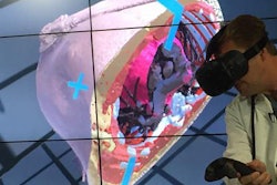
A little time spent learning about 3D, maximum intensity projections, and cinematic rendering could be well spent. Cinematic rendering is a photorealistic 3D rendering technique that enables a more in-depth 3D evaluation of the underlying anatomy, presenters say.
There's even a session on using today's tools optimally to make your and your patients' lives better while dealing with a deluge of images. And isn't that what it's all about?
Learn more about these sessions in the previews below. To view RSNA's complete listing of abstracts for this year's scientific and educational program, click here.