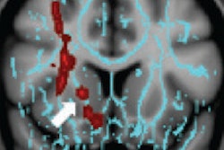
Using diffusion-tensor MRI (DTI-MRI), researchers have found differences in brain wiring between children with sensory processing disorders (SPD) and those with autism, according to a study published online July 30 in PLOS One.
The group from the University of California, San Francisco (UCSF) found that children with SPD had decreased structural brain connections in specific sensory regions different from autism, further establishing SPD as a clinically important neurodevelopmental disorder.
This new research follows a 2013 UCSF study that suggested boys with SPD had quantifiable regional differences in brain structure when compared with typically developing boys.
The current study examined the structural connectivity of specific white-matter tracts in 16 boys with SPD and 15 boys with autism between the ages of 8 and 12 years. The researchers compared the findings with those of 23 typically developing boys of the same age range, noted senior author Dr. Pratik Mukherjee, PhD, and colleagues.
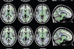
Both the SPD and autism groups showed decreased connectivity in multiple parieto-occipital tracts, which handle basic sensory information. However, only the autism cohort showed impairment in the inferior fronto-occipital fasciculi, inferior longitudinal fasciculi, fusiform-amygdala, and the fusiform-hippocampus tracts, which are critical for social-emotional processing.
Kids with isolated SPD showed less connectivity in the basic perception and integration tracts of the brain that serve as connections for the auditory, visual, and tactile systems involved in sensory processing.