With an eye toward improving the early diagnosis of neurological disorders such as Alzheimer's disease, GE Healthcare this week revealed that it is developing a compact 3-tesla MRI scanner designed specifically for head and brain imaging.
The company is collaborating with the Mayo Clinic in a public-private partnership funded, in part, by the U.S. National Institutes of Health (NIH). The short-term goal is to test the technology in a clinical environment at the Mayo Clinic as soon as early next year.
"We are trying to understand how a small, dedicated system for head imaging can fit in the clinical environment and what benefits it can provide," said Luca Marinelli, PhD, manager of GE's MR research laboratory in Niskayuna, NY.
The brain MRI prototype is significantly smaller in size and weight than a conventional whole-body MRI scanner. GE researchers have a target weight of approximately 2,000 lb for the system, which would allow the device to be located on any floor within a hospital or imaging center with no need for structural reinforcement. It would also have a considerably smaller footprint than a whole-body scanner.
The prototype's gradient coil design is made to accommodate the human head, with an anticipated bore size of approximately 38 cm when the device is completely assembled. As a patient's head glides into the scanner, his or her shoulders would bump up against the edges of the bore to prevent the person from going too far into tube.
"The imaging field will be limited to the head down to the top of the cervical spine," Marinelli explained.
Shorter scans
Early estimates are that the compact system could reduce scanning time to approximately 10 minutes, which would be especially beneficial for patients in pain or who may be unable to spend a great deal of time in a conventional MRI scanner for whatever reason.
Another feature of the compact system is that it would require a much smaller amount of liquid helium for cooling during operation.
"Today, a conventional MRI scanner needs approximately 2,000 liters of liquid helium, which needs to be periodically refilled," Marinelli said. "The new system features closed-loop refrigeration and would use only 12 liters of liquid helium."
Getting access to large amounts of liquid helium has become "an extreme constraint" in recent years, Marinelli added, due to constant shortage and supply issues.
So far, so good
"We know the magnet works with the no-heating technology," said Nadeem Ishaque, GE's global technology director for diagnostics and biomedical technologies. "The [gradient] coil is a very special coil which required a lot of engineering; we know this works. We still have to put the two together to make sure the whole system works."
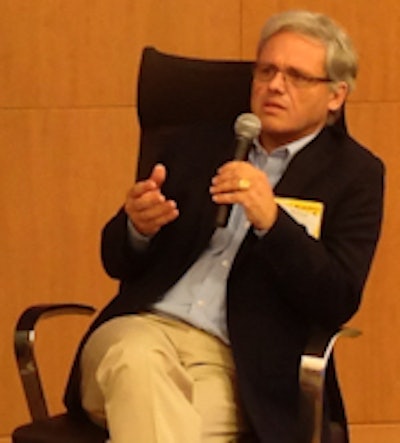
 Dr. Patrick Hof from the Icahn School of Medicine at Mount Sinai addresses attendees at the GE Research Center on the state of brain imaging.
GE touted the brain-imaging MRI prototype this week during a tour of its research facility in Niskayuna. The attendees included Dr. Patrick Hof, vice chair of the department of neuroscience at the Icahn School of Medicine at Mount Sinai.
The role of PET and MRI in brain imaging is becoming increasingly important to the diagnosis of degenerative brain disorders with the emergence of a number of PET ligands principally centered around amyloid pathology and tau markers, he said.
"The combination of PET and MRI is very important because we can get functional images and we can map them," Hof said. "As both technologies get better and better in terms of resolution, ultimately, we can dream of getting your cellular resolution as a routine."
Hof also expects to see more biomarkers "for whole families of cells, like microphages, that could eventually be used in combination with functional imaging. So good PET/MRI systems with optimized readouts will be extremely important to assess the affected populations," he said.
This area of neurological imaging research also will open an entirely new branch of diagnostic radiology, which specialists and researchers "will have to grow into and adapt to," he added. "That will involve the formation of fellows and residents to these novel techniques, which currently does not exist."
Expanding applications
GE is also implementing a number of applications and techniques for neuroimaging that have already proved useful on its full-size MRI scanners, such as the Discovery MR750.
Among its research projects, the company is collaborating with the University of California, San Francisco (UCSF) on a resting-state functional MRI study to assess the brain's network and how regions work together in sync. Subjects are imaged while lying passively in a resting state, rather than being scanned while performing a task.
"We look at the whole brain simultaneously and then use postprocessing techniques to extract correlations that allow us to determine what part of the brain is related to motion, what part of the brain is related to attention, and what part of the brain is doing nothing or is the default mode network," Marinelli said.
GE also recently began investigating fundamental magnetism within the brain at its research center. The accumulation of magnetic materials in the brain, such as iron, occurs both with normal aging and in certain regions, which could indicate disease. It is well-known, Marinelli said, that iron accumulation in the substantia nigra region is associated with Parkinson's disease.
MRI's promise
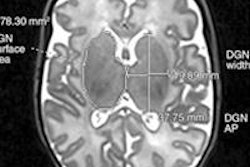
In addition, traumatic brain injuries can cause microbleeds in the brain, which can be difficult to detect. Brain MRI could help find microbleeds that otherwise might not be discovered.
"Microbleeds and calcifications with conventional [imaging] techniques look exactly the same," Marinelli said. "Microbleeds and calcifications from the perspective of magnetic properties of the brain are on opposite ends. Blood is paramagnetic, while calcifications are diamagnetic to water. So one enhances the magnetism and one diminishes the magnetism of the brain."
Even with substantial advances in recent years, there is much progress to come with MRI technology.
"We are working on the next generation of MRI where we believe we can take down the resolution to tens of microns," Ishaque said. "This is all research; it will not be a product anytime soon, but with fundamental research we can start to look at the architecture of the brain in terms of what the cells are, completely in vivo, completely noninvasive without a contrast. That will be the dream."