It is well known that medical radiation dose is greatly affected by patient size. Less well known, however, is the impact of other factors, such as fat composition. Authors of a new study in Physics in Medicine and Biology got a better handle on dose by creating new phantoms that more closely approximate the body habitus of overweight patients.
Obesity has become a significant public health problem in recent decades, with 60% of the U.S. population now defined as clinically overweight or obese. Excess weight is also linked to a number of diseases, such as cardiovascular disease, type 2 diabetes, and cancer. As a result, obese patients represent an increasing proportion of all patients scanned. But there is little information about the kinds of radiation doses these patients are receiving when they undergo scans.
But although obesity is known to have profound effects on CT image quality and patient organ dose, quantitative data describing this relationship have not been available, wrote Aiping Ding, PhD, Matthew Mille, and colleagues (Phys Med Biol, April 6, 2012).
"The goal of our study was to develop a new set of obese phantoms to represent obese patients," said lead author Ding in an interview with AuntMinnie.com. "Second, we wanted to study how much radiation dose overweight patients receive from CT scans, and third, we wanted to compare dose readouts between normal-body-weight and obese patients."
The results were sometimes surprising.
"We found that obese patients receive very high doses -- higher than we thought before we performed this study," Ding said. "We were surprised to see the dose readouts."
Building fatter phantoms
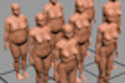
The investigators from Rensselaer Polytechnic Institute (RPI) in Troy, NY, built 10 of their own special "obese phantoms" to simulate radiation dose effect for larger individuals. The 10 phantoms (five males, five females) had body mass indexes ranging from 23.5 (normal body weight) to 46.4 kg m-2 (morbidly obese). Incorporating more than 100 deformable organs defined by mesh geometry, they were derived from earlier adult male and female computational phantoms developed at RPI. The phantoms included measured amounts of subcutaneous adipose and visceral adipose tissue.
The phantoms were then voxelized and defined within the Monte Carlo N-Particle Extended code to calculate organ doses from CT imaging, and their weight, height, and organ size of RPI-AM (male) and RPI-AF (female) were carefully adjusted to match with reference values defined by the International Commission on Radiation Protection (ICRP) for average individuals, Ding and colleagues wrote. The modeling procedure assumed that organ mass remains constant during adulthood, which is "a decent approximation for the purpose of radiation protection dosimetry," that has been used by others, they wrote.
The results showed that for obese patients, radiation doses to organs deep in the abdomen, such as the colon, are reduced by more than half compared to those for normal-weight individuals, while shallow organs received higher doses.
"Going from normal to morbidly obese, the fatty tissues increased, especially in the abdominal region, so if we use the same CT settings to scan normal-weight and obese patients, the dose to deep organs decreases significantly, but the dose to superficial organs didn't change much because there's less fatty tissue," Ding said.
Increasing the tube current -- standard practice for scanning larger patients -- raised the organ doses by well over half in amounts that rose with the level of obesity, the study found. Most scanners aren't designed for large patients, but there are large-bore scanners that are sometimes used in extreme cases to accommodate patients too large for the approximately 70-cm port diameters typical of most commercial CT machines.
Protocols need to be adapted to fit these patients; in clinical practice with CT, that typically means increasing the field of view, the tube potential, and the tube current to accommodate them, which results in generally higher doses for these patients, the authors stated.
Different kinds of fat
But it's not just the amount of fat that affects dose and image quality, but the type of fat, Ding said. Adipose tissues can be divided into two classes: subcutaneous adipose tissue (SAT), which is located beneath the skin, and visceral adipose tissue (VAT), which surrounds the abdominal organs, he said.
"As discussed in previous studies ... VAT is more metabolically active than SAT, and of the two categories of fat, it is the most strongly correlated with many of the negative health effects often associated with obesity," the authors wrote.
VAT is a major risk factor for obesity-related diseases, and it plays an important role in the effect of adipose tissues on the radiation dose delivered to the deep organs of the abdomen. But because little is known about how organs move or deform in the presence of excess VAT, "the same region inside the peritoneal cavity was used to define the VAT volume for all phantoms, regardless of their BMI values," the authors explained.
Scanner settings were based on previously developed protocols for a LightSpeed 16 scanner (GE Healthcare) consisting of 16 rows of 0.625-mm-wide detectors and 8 rows of 1.25-mm detectors. The study was based collimation of 20 mm, 120-kVp and 140-kVp tube potentials in normal-weight, overweight, and obese phantoms, and a pitch of 1.375.
As the effect of adipose tissue on CT imaging dose will be the largest in the torso of the patient, a chest-abdomen-pelvis (CAP) scanning protocol was simulated for each of the BMI-adjustable phantoms. The scan range was chosen to cover the phantoms from just above the clavicles to the mid-iliac crest. The scan lengths for the RPI BMI-adjustable male and female phantoms were 44 cm and 41 cm, respectively.
Because the same tube potential (120 kVp) and current time (100 mAs) were used for all 10 phantoms, CT radiation doses to superficial organs such as the breasts for all the different BMI value phantoms were found to decrease slightly as BMI increased. But organs deep within the body (including stomach, gonads, urinary bladders, liver) were highly shielded by VAT and/or SAT, and as a result, the dose values for these organs decreased significantly with increasing BMI.
Average skin dose falls, cumulative skin dose rises
As skin mass grew with increasing BMI values, the average skin dose fell between 7% and 13% in the male phantoms and between 4% and 11% in the female phantoms. Nevertheless, once the differences in skin mass were accounted for, the total energy deposited in the skin was found to increase with BMI by factors of 1.07-1.28 in the male and 1.02-1.19 in the female phantoms.
"This demonstrates that obese patients receive a higher skin exposure because the distance between the skin and CT x-ray source decreases," Ding and colleagues wrote.
Of course, using the same kVp and mAs isn't realistic for clinical practice, because the thicker body dimensions of overweight and obese patients must be compensated for by greater tube current or higher tube potential in order to maintain acceptable image quality, the study team wrote.
The higher tube potential increases x-ray output, but the ways in which it does so are not always readily predictable. According to the authors, "the relationship between the radiation dose, image noise, and kVp setting is quite complex, and the proper selection of kVp depends on the desired image quality for a given examination." As a result, higher tube current increases the organ doses.
When tube potential was increased from 120 kVp to 140 kVp for the obese phantoms, keeping the mAs constant, the organ doses for the morbidly obese RPI phantom increased by as much as 62% (male) and 59% (female).
Nevertheless, organ doses for organs deep in the abdominal region (including colon, stomach, gonads, urinary bladder, and liver) were found to be lower for the morbidly obese phantom compared to the normal-weight phantom when a 120-kVp tube potential was used for both phantoms. These deeper organs are heavily shielded by the extra adipose tissue, especially by the VAT, Ding et al explained.
When the concept of VAT shielding was further validated by replacing the VAT in obese individuals with normal body tissue and repeating the same scans, the dose to the deep organs grew by as much as much as 16% (male) and 32% (female) for the morbidly obese phantoms due to reduced organ shielding. The results show that VAT should absolutely be taken into consideration when calculating radiation dose for obese individuals, they wrote. This effect was less significant for shallow organs such as the skin and breasts.
"It was also found that if VATs were ignored in the modeling process of obese individuals (i.e. VAT replaced by normal body tissues), the dose to organs deep in the abdominal region increased by as much as 16% and 32% in the obese male and female phantoms, respectively," the authors concluded. "The results from our simple VAT model suggest that VAT plays an important role in the organ dose estimates and should always be considered for obese individuals. As expected, the data confirm that an increase in kVp and mAs results in an increased absorbed organ dose and effective dose to the obese patients."
When scanning an obese patient "the radiologist needs to increase the power setting to generate higher energy and get better images," Ding said. But until now the process has been accomplished mostly by "trial and error," without scientific models, he said.
Later this year, the group will incorporate the models into a software package that can be used to guide the scanning of all types of patients, from children to pregnant patients to the morbidly obese. It will show how much noise increases for a given protocol in a given patient type, and it will include radiation dose levels both inside and outside the scan field, Ding said. The obese phantoms created for this study can also be used for future research in image quality and organ doses in CT, as well as PET, SPECT, and interventional radiography, the group noted.
The study was funded primarily by the U.S. National Institutes of Health (NIH), with additional support from the National Institute of Biomedical Imaging and Bioengineering (NIBIB), the U.S. Department of Energy's (DOE) Office of Nuclear Energy's Nuclear Energy University Programs (NEUP), and the Health Physics Society.