LONDON - An exhibition that highlighted the value of CT to reveal millennia-old secrets of mummies from ancient Egypt and Sudan drew to a close recently at the British Museum. But such was the popularity of "Ancient Lives, New Discoveries" that its run was extended twice, and now plans are being made to take it on an international tour toward the end of 2016.
The ancient lives portrayed in the exhibition date back to 3,500 B.C. and include both high- and low-status inhabitants of ancient regions of Gebelein, Thebes, Hawara, and et-Tereif. Discoveries made with single- and dual-energy CT included dental abscesses, atherosclerosis, and novel insights into burial rituals and practices at the time.
"In some respects, it was a test case exhibition for us to see if we could mount a show like this focusing just on human remains and present it in a respectful and sensitive way, and avoid anything sensational and ghoulish," said Dr. John Taylor, curator on ancient Egypt (funerary archaeology, ancient Egypt and Sudan). He spoke at "The Curious World of Radiology," a seminar organized last week in London by the British Institute of Radiology (BIR).
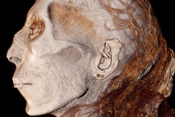
 This mummified man was buried in a crouching position around 3,500 B.C. in Egypt and was discovered in 1896. Thought to be between 18 and 21 years old when he suffered a violent death, he was wrapped in linen and matting and placed in a shallow grave. He was stabbed in the back by a weapon. All images of mummies courtesy of Trustees of the British Museum.
Due to the perils of unwrapping mummies, which can lead to disintegration of the bodies, CT was used to look inside the mummies without causing damage. CT scanning has reached a level of such sophistication that we can pinpoint incredible detail inside the mummies, turning CT into 3D volumes and then segmenting the data, he told attendees. Eight mummies were scanned for the show, but the project is ongoing, and Taylor and his colleagues have recently scanned a further six mummies that they would like to add into the potential touring version of the exhibition.
"With our new work, are looking for similar stories to those seen with the original set of mummies, but we seek new information too," he remarked. "We'd like to focus on life expectancy, health and nutrition, pathological conditions, as well as the process of mummification."
The original exhibition revealed there was a lot more variation than was expected in the rituals and processes practiced for mummification. Standard textbooks espouse three main methods of mummification, and it has been common belief that embalmers in ancient Egypt carefully followed these, but CT scanning is changing the way historians are thinking about this.
"We are now finding that there was a much wider spectrum of techniques and also things didn't always go to plan," said Taylor, adding he was surprised to find damage to the bodies carried out by the embalmer, which has refreshed knowledge on how the ancient Egyptians viewed the human body. "From a view that keeping the body intact was paramount, it appears that they were actually prepared to have it disarticulated as long as it was put back together again, and the outside looked good."
In very early Egyptian civilization, this disarticulation may have been an aid to preservation of the soft tissues in particular. They may have opened up the body and deliberately allowed the soft tissue to decompose and then the bones were reassembled, Taylor continued. Also, there may have been a religious rationale to support this process. Disarticulating and reassembling the body may have been reminiscent of a myth about the god of the dead, Osiris, which simply put, says that on death, a body was cut to pieces and then reassembled.
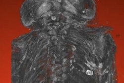
 Image shows use of sticks to reassemble the head with the body during mummification.
"The head of one mummy had completely snapped off, and the embalmer pushed it back on with wooden poles," he said. "We can tell by looking at the neck vertebrae that this had happened postmortem during the drying process. The sticks were part of a running repair."
Pathological evidence
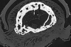
Large, calcified plaque deposits, effectively evidence of atherosclerosis was found in two of the femoral arteries of two of the mummies. The high status of most of the mummies might have meant they ate high-fat diets, or there may even have been a genetic factor at play. Using CT, much more of this type of evidence can now be determined, according to Taylor, who hopes to see more evidence of disease, including dental disease.
"Could these dental problems have been fatal to these people? One of our big questions is what was the cause of death of these mummies? It has been suggested that some of the dental abscesses were so severe that they may have died from blood poisoning," he stated.
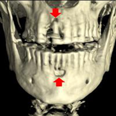
 Image shows location of possible dental abscesses.
Some mummies are so-called natural mummies -- bodies that were simply buried in the sand and preserved by the dryness of the environment. These mummies have their internal organs intact and differ from high-status mummies whose organs were removed and kept in jars, but often little tissue is well-preserved this way.
"Natural ones are more interesting because they are very well-preserved," Taylor noted. "One of our studies now is to look at more natural mummies. These are very early bodies that are around 5,500 years of age, and should provide good insight into the population biology of Egypt at the time."
 Details about new technical developments and innovations that can be applied to examining mummies would be valuable, Dr. John Taylor said. Image courtesy of BIR.
He also hopes to find more information on amulets, which are small objects often found in the shape of animals or plants and thought by ancient Egyptians to protect the body on its journey through the underworld.
"Unfortunately, although many amulets have been found previously in mummies, very few were documented, and we do not know the positions these amulets occupied. The value of scanning is that we see them in their correct locations," he said.
Some amulets have inscriptions on them, and Taylor hopes that CT imaging may soon reach the point where these inscriptions will be visible. An amulet inside the mummy that is visible on CT imaging could reveal a person's name, he believes.
Finally, in a plea to radiologists, he pointed out that he and his colleagues are always interested in new technical developments and innovations that might be applied to examining mummies.
"We look to the radiology community for any leads on this front," Taylor concluded.
Further information about the exhibition can be read in AuntMinnieEurope.com's article on it last year, as well as on the British Museum's dedicated website.