A modified CT enteroclysis (CTE) technique, performed with widely available and affordable infusion equipment and enteral contrast, can obtain diagnostic-quality images of small-bowel distention, South African researchers have reported in the European Journal of Radiology.
"This is of particular value in developing countries where we are resource-limited and expensive equipment and contrast material is often not available," noted lead author Dr. Braham van der Merwe, a radiology registrar at Tygerberg Hospital, University of Stellenbosch, Cape Town. "Experience and evidence-based analysis have shown that only small-bowel enteral volume-challenged examinations that uniformly distend the small-bowel lumen can confidently confirm or rule out small-bowel pathology."
 CT enteroclysis (CTE) axial and coronal images of a patient with Crohn's disease. Axial image demonstrates active inflammatory changes of terminal ileum with hyper enhancement. An added benefit of the South African authors' CTE technique is that it's possible to fill the colon with negative contrast, as can be demonstrated on the coronal image with active inflammatory changes of the cecum and ascending colon. All images courtesy of Dr. Braham van der Merwe.
The small intestine is the most challenging segment of the gastrointestinal tract to examine and image because it has the largest mucosal surface. Incidence of disease is low and clinical presentation is mimicked by adjacent visceral organs with higher incidence of abnormalities, the authors explained in an in-press article posted by EJR on 8 May 2013. Thus imaging of small-bowel disease remains poorly understood by referring clinicians and radiologists.
CTE was introduced at Tygerberg Hospital in July 2010. All adult patients (73) undergoing CTE between July 2010 and November 2011 were included in the study. Four failed duodenal intubations had received CT enterography, so they were excluded.
Of the 73 patients, 42 (57.5%) were female, and the mean age was 50.1 years and a standard deviation (SD) of 16.8 years. The study group included 39.7% (29) Caucasians, 11% (eight) black, and 49.3% (36) colored (mixed ethnic origin) patients. No statistically significant associations between nominal data, race, or sex were found in the relevant subgroups.
A total of 15 (20.5%) of the 73 patients had positive findings on CTE related to small-bowel pathology, and 12 (16.4%) had nonsmall-bowel pathology that could explain clinical symptoms. Most prevalent small-bowel findings were in the inflammatory bowel subgroups (either known or suspected), where six (30%) patients out of 20 had signs of active inflammatory bowel disease (IBD).
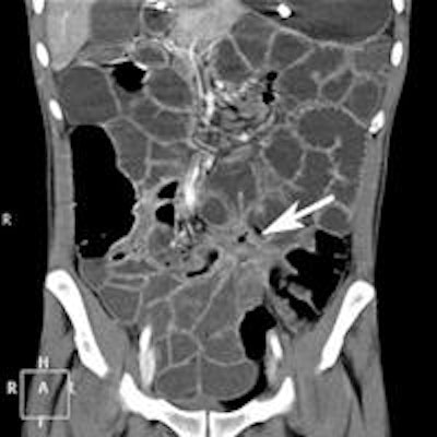
 CTE coronal image of a patient with Crohn's disease. Value of CTE due to volume challenge is illustrated with fibrostenosing disease with fistulation. This can easily be missed on a nonvolume-challenged study.
Based on the indications for referral, the largest group was the malabsorption/chronic diarrhea group with 19 (26.03%) referrals, according to van der Merwe and colleagues. In this group, one patient had celiac disease, one had Crohn's disease, and one had ulcerative colitis with terminal ileum involvement. The anemia group was the second largest with 13 (17.81%) referrals. In this group, no intestinal cause was identified for anemia. One patient had multiple uterine fibroids.
In the group of patients with known IBD, 3 (37.5% of subgroup) had signs of active inflammation and one patient had perianal fistulae. In the group referred with possible IBD, 3 (25% of subgroup) patients had signs of active IBD, one had ileocecal intussusception and one had a small-bowel mass lesion. This proved to be a hamartoma at histology following surgical resection.
"With our new and improved method using saline, drip sets, and a three-way tap, we are able to achieve uninterrupted bowel distention. This allows for an easy, reproducible, and effective way of performing CTE, with adequate distention thus a more reliable way of excluding small-bowel abnormalities," the authors stated.