Because it's noninvasive and doesn't require anesthesia, CT postprocessing offers significant advantages over the gold standard of bronchoscopy for evaluating pediatric airways. Yet there are even more benefits, according to an article published recently in Pediatric Radiology.
CT with postprocessing for evaluating children's airways provides pediatricians with imaging perspectives that match their clinical and anatomical understanding, allowing them to make better decisions from the imaging information, lead author Dr. Savvas Andronikou, from University of the Witwatersrand in South Africa, told AuntMinnie.com.
"It helps pediatricians understand what a radiologist is reporting and allows them an opportunity to show parents better what affects their children," he said.
Andronikou and colleagues discussed technical developments in postprocessing of pediatric airway imaging in a recent minisymposium article (Pediatr Radiol, March 2013, Vol. 43:3, pp. 269-284).
In contrast to bronchoscopy, CT does not require anesthesia or the passage of a tube into the airway, he said.
"CT is not without risks -- the radiation burden is far greater than plain x-ray -- but there is no discomfort or invasion of the child and reconstructions are performed from already available CT information," he said.
Furthermore, bronchoscopy can only evaluate the internal aspect of the airway and is prevented from further passage at narrow parts of the bronchial tree.
"CT can see beyond narrow areas, and, in addition, one can see outside the airway and determine the cause of a narrowing, being able to distinguish masses from blood vessels, accurately," he said. "Bronchoscopy does allow some interventions such as biopsy and suction that help with diagnosis or represent therapeutic measures, but this invasive procedure can be reserved for patients where this is thought necessary, after reviewing of CT scanning."
It's also important to note that reconstructions are performed from standard MDCT scans without any additional or special scanning, Andronikou said.
"All types of postprocessing reconstructions can be achieved from the same set of CT images, and these can be performed over and over, even on freeware viewing platforms on personal computers," he said.
Postprocessing tools
A range of visualization techniques can be called upon at clinical workstations, such as multiplanar reformation (MPR), including curved planar reformation (CPR); maximum and minimum intensity projection (MIP, MinIP); shaded surface display (SSD); and volume rendering (VR).
VR is the most accurate CT reconstruction tool, but it's user-dependent and mostly used by radiologists, according to Andronikou. MinIPs are ideally suited for imaging children's airways "because they represent a slab of tissue including and preferentially representing air-containing structures in their full thickness," he said.
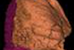
Another technique, endoluminal virtual bronchoscopy, can be created by both SSD and VR and mimics fiber-optic bronchoscopy. This provides pediatric pulmonologists with a CT scanning view they are familiar with, Andronikou said.
In future developments, computer-aided diagnosis (CAD) may also find a role in pediatric airway imaging, with recent research showing promise for CAD in diagnosing tuberculosis.
Radiologists routinely use CT reconstructions, particularly MPR, as part of standard viewing for diagnosis, he said.
More complex reconstructions are currently performed by radiologists at workstations and are user-dependent and time-consuming, Andronikou said. Furthermore, there are no standard recommendations for storing these created images.
Expanding clinician use
For more widespread clinician appreciation and use of CT postprocessing, radiologists need to provide a standard set of reconstructions for particular indications.
"In pediatric chest imaging, a useful standard reconstruction that should be supplied is a thick-slab MinIP of the airway in the coronal plane," he said. "This can be produced as a standard and supplied with the routine axial images."
Of course, pediatricians then need to understand what limitations exist and what is being demonstrated accurately in order to gain confidence in the tool, he said.
Radiologists could also routinely supply a video clip of virtual bronchoscopy oriented in a standard way that mimics true bronchoscopy.
The alternative would be to train pediatricians to produce these images on their personal computers using freeware such as the OsiriX image viewing application.
Postprocessing techniques can demonstrate fine anatomical detail and provide additional information that is relevant for diagnosing and managing airway disease, the authors concluded. These tools also supply images that are easy to interpret, specifically for improved communication with referring clinicians and parents.
In addition, postprocessing methods have potential for automating airway visualization, identifying airway anatomy, and detecting regions of pathology, according to the group.
"Improved airway segmentation will offer improved visualization of the smaller bronchi and allow automatic extraction of the airway structure," the authors wrote. "Branch measurements can then be automated and analysis of large datasets of images can be performed. These methods would allow clinicians to view the airways with overlays indicating airway properties or the likelihood of disease based on comparison to a dataset of airway images."
