CHICAGO - MRI is generally the go-to modality for differentiating old and new vertebral fractures, but if a magnet is out-of-pocket, then multidetector-row (MDCT) can be used instead, according to a presentation this week at the 2007 RSNA meeting.
Dr. Ji Young Hwang and colleagues from Ewha Woman's University School of Medicine in Seoul, South Korea, evaluated the value of MDCT in discriminating acute vertebral fractures from old vertebral fractures.
"Acute vertebral fractures are common and occur because of trauma or osteoporosis. Determining the stage of a fracture is critical for performing vertebroplasty and for (insurance payment)," Hwang said. "Currently, MRI is used to determine the stage of a fracture. Because MDCT is easily available, it is the initial diagnostic tool for vertebral fracture. However, the diagnosis accuracy of MDCT in determining the stage of fracture has not been determined."
The study population consisted of 173 vertebral segments in 122 consecutive patients with acute vertebral fractures. The most common cause of injury was a fall from a great height followed by motor vehicle accident. The majority of the patients had compression fractures at the T12-L2 level.
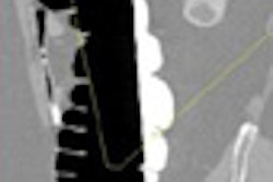
Patients underwent MDCT exams with a protocol that included axial (2-mm slice thickness) and sagittal (3-mm slice thickness) multiplanar reconstruction (MPR) imaging. The patients also had MR studies and the protocol included T1-weighted sequences, as well as T2-weighted fat-suppressed sequences. The MR studies were used as the reference standard, Hwang said.
Two musculoskeletal radiologists reviewed the MDCT images in consensus, looking for the presence of fracture line, loss of paravertebral fat plane, and changes in marrow density relative to adjacent normal vertebrae.
According to the MR results, 144 (83%) of the vertebral segments had acute fractures and 29 (16.7%) had old fractures. In 92% of the cases, fracture lines were seen on MRI, as were changes in marrow signal intensity (100%). In comparison, MDCT results led to the correct diagnosis of fractures in 147 cases (85%). On MDCT, fracture lines were found in 129 (74%) of the cases, while loss of paravertebral fat plane was noted in 125 (72%). Change of bone marrow density was seen in 119 (69%) of the cases. MRI did spot spinal cord injury in two cases and 10 instances of ligament injury.
An RSNA session attendee asked Hwang about the frequency of cord injuries that were seen on MRI but not on MDCT. Hwang replied that in this patient population, there were two cord injuries, but both were seen on MDCT scans.
Hwang reported that for all findings, MDCT turned in a sensitivity of 94%, a specificity of 69%, and an accuracy of 90%. Her group concluded that their results demonstrated the key signs of acute vertebral fracture on MDCT. In particular, radiologists should look for subtle fracture lines on the sagittal images, changes in marrow density, and paravertebral fluid collection, she explained.
By Shalmali Pal
AuntMinnie.com staff writer
November 29, 2007
Related Reading
Axial-loaded imaging of lumbar spine heightens diagnostic accuracy, May 29, 2007
Spinal Chance-type fractures require careful imaging analysis, November 9, 2006
CT more sensitive than spinal fracture x-ray, but has limits, January 30, 2006
MRI sees spinal metastases earlier, but low-dose whole-body MDCT aces skeletal workup, May 20, 2005
Copyright © 2007 AuntMinnie.com