CHICAGO - Computer-aided detection (CAD) technology offers promise for improving radiologists' performance in assessing the likelihood of lung nodule malignancy, according to research from the University of Michigan Medical School in Ann Arbor.
"CAD has the potential to serve as a second opinion for lung nodule characterization," said Ted Way. He presented the study team's findings Sunday at this week's RSNA 2007 meeting.
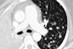
To evaluate the effect of CAD on radiologists' estimates of malignancy of lung nodules on CT, the researchers performed a receiver operating characteristic (ROC) study using their CAD system. They included 152 patient CT scans containing 256 nodules, including 124 malignant and 132 benign nodules. Pathology was established using biopsy or two-year follow-up.
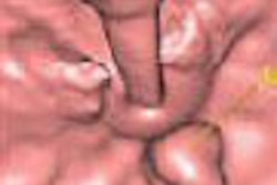
Six thoracic, fellowship-trained radiologists served as observers. Following a training session for each observer to familiarize them with the user interface employed in the study, the radiologists read the exams sequentially, first without CAD, then immediately following with CAD. Observers had the option of utilizing 3D rendering in their review process, according to Way.
They rated the likelihood of malignancy using a 0 to 100 scale. Nodules from the same cases were separated, and a different randomized order was used for each observer. There was no time limit, and observers could complete the study in one or multiple sessions, Way said.
The data was then analyzed using the Dorfman-Berbaum-Metz method, as well as with LABMRMC software.
The CAD system achieved a test Az of 0.86 using three morphological, two texture, and two gradient field features. All six radiologists were found to have improved performance with CAD, with four reaching statistical significance (p < 0.05). Average Az of the radiologists improved significantly from 0.83 without CAD to 0.85 with CAD.
In examining the effect of reading with CAD, the researchers found that malignancy assessment was changed in 126 of the 256 nodules. CAD led to an improved assessment in 95 of those cases. A change in recommended action was found in 11 of the 256 nodules, of which seven were an improvement, Way said.
"We did see a modest, though significant improvement," he noted.
One reason for the modest improvement could be the use of thoracic, fellowship-trained radiologists in the study, Way said.
"Maybe (with) general radiologists or radiologist residents we might see different results," he said.
In one case involving non-small cell lung cancer, the six radiologists had an average classification of 45.8 (range of 5-70) without CAD. Following CAD results, however, that classification increased to 58.3, Way said.
In a benign nodule case, radiologists had an average classification of 53.3 (range of 20-65). Following CAD results, however, that rating decreased to 48.3, he said.
In one benign case that was biopsied and found negative for neoplasm, radiologists had an average classification of 73.3 (range of 40-90). Following CAD results that incorrectly judged the nodule to be more likely malignant, that score only decreased to 68.3.
"This showed that radiologists can still independently assess the results of CAD," Way said.
The radiologists' ability to assess malignancy was improved significantly with CAD, he concluded.
"All radiologists showed individual improvement," Way said.
By Erik L. Ridley
AuntMinnie.com staff writer
November 25, 2007
Related Reading
CARS news: USC group develops home-grown CAD-PACS integration toolkit, June 29, 2007
Divergent research on CT lung screening sparks more debate, fewer answers, April 19, 2007
CAD performs well in lung nodule detection, February 5, 2007
Lung CT CAD boosts performance of less experienced radiologists, November 27, 2006
Copyright © 2007 AuntMinnie.com