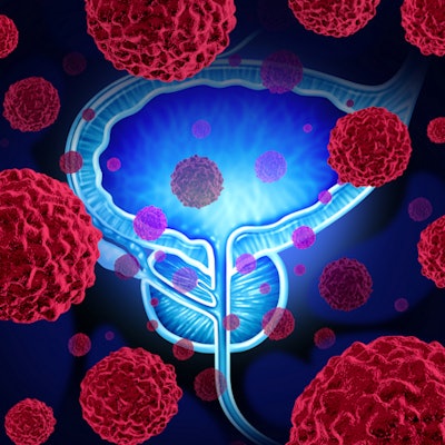
MRI scans with a reduced dose of a gadolinium-based contrast agent (GBCA) can be more effective than scans with a standard dose in differentiating between malignant and benign lesions in prostate cancer patients, according to a study presented recently at RSNA 2017.
A lower GBCA dose resulted in significantly greater signal enhancement in patients with malignant tumors than in those with benign prostate tissue. In comparison, there was no significant difference in signal when patients were given a standard dose of a GBCA.
"Dynamic contrast-enhanced MRI is important for prostate cancer detection," said lead author Dianning He, a research student at the University of Chicago. "Quantitative dynamic contrast-enhanced MRI with low gadolinium dose better distinguishes prostate cancer from benign prostate tissue than standard gadolinium dose, based on signal enhancement rate."
GBCA debate
The use of gadolinium contrast for all types of MRI scans has come under closer scrutiny in recent years following studies that showed minute traces of gadolinium in patients long after they received contrast. Gadolinium deposition predominantly has been discovered in certain regions of the brain based on increased signal intensity during unenhanced MRI scans.
The jury is still out on whether there are long-term adverse effects from gadolinium deposition. While several studies have found no conclusive evidence of harm, many patients blame a range of ailments on having been given gadolinium for routine MRI scans.
Although He and colleagues did not study the potential effects of gadolinium in the prostate, he cited recent studies that recommended lower doses of GBCAs when possible.
"In this study, we investigated whether the administration of low-dose gadolinium for prostate MRI scans is as effective as a standard dose for the detection of cancer and prostate benign tissue," he told attendees at RSNA 2017.
The study included 17 patients with histologically confirmed prostate cancer. They underwent preoperative 3-tesla MRI scans using endorectal and phased-array surface coils. GBCA-enhanced MR images were acquired using two mDixon sequences with a temporal resolution of 8.3 seconds. The lower contrast dose used was 0.015 mmol/kg, while the standard dose was 0.085 mmol/kg. The patients received gadobenate dimeglumine (MultiHance, Bracco Imaging), a linear GBCA, in the scans.
The protocol included the acquisition of MR images with the low-dose GBCA protocol over 3.5 minutes. Five minutes after completing the low-dose scans, clinicians acquired standard-dose images for 8.3 minutes.
He and colleagues later analyzed signal intensity using a mathematical model to calculate maximum intensity projection and signal enhancement rate. A receiver operating characteristic (ROC) analysis was performed to evaluate how each protocol affected radiologists trying to distinguish between prostate cancer and benign tissue. Prostatectomy specimens were used as the reference standard.
By the numbers
Most importantly, the researchers found that low-dose GBCA scans produced higher signal enhancement rates for malignant prostate tissue than standard-dose GBCA. There was no statistically significant difference in signal enhancement rates between malignant and benign tissue when the standard dose of GBCA was used.
| MRI with low- vs. standard-dose GBCA for prostate cancer | ||
| GBCA dose | Signal enhancement rate | |
| Prostate cancer | Benign tissue | |
| Low | 10.0 (± 5.8) | 5.1 (± 2.9) |
| Standard* | 4.3 (± 2.2) | 3.4 (± 1.5) |
The ratio of low-dose to high-dose signal enhancement was significantly greater for patients with prostate cancer (2.8 ± 2.3) than for those with normal tissue (1.6 ± 0.9). He and colleagues suggested that the greater signal enhancement from a low dose of GBCA was due to T2-weighted effects associated with cancer.
In addition, the area under the ROC curve for differentiating prostate cancer from benign tissue using signal enhancement was higher for low-dose GBCA (0.77, p < 0.05) than the standard dose (0.63, p > 0.05).
As for the maximum intensity projection parameter, the researchers found no significant differences for prostate cancer and normal tissue at both low (33 ±12% vs. 29 ± 16%) and standard doses (110 ± 49% vs. 94 ± 43%).
He cited several limitations to the study, including the low number of subjects and the fact that residual contrast from administration of the low dose could interfere with the analysis of the standard dose.
"Additional work is needed to determine the dose and the injections that are optimal for prostate cancer diagnoses," he said.