MRI brain scans taken before and after the start of football season have uncovered abnormalities in young players' default mode network after just one season, according to two studies presented on Monday at RSNA 2017 in Chicago.
The brain's default mode network engages when people are awake and helps with the processing of emotions. The new findings add to growing evidence that subconcussive head impacts can adversely affect the brain, especially among young athletes.
 Gowtham Krishnan Murugesan from UT Southwestern.
"Our results suggest an increasing functional change in the brain with increasing head impact exposure," said Gowtham Krishnan Murugesan, a doctoral student in biomedical engineering at the University of Texas (UT) Southwestern Medical Center, in a release from the RSNA. "This study demonstrates that playing a season of contact sports at the youth level can produce neuroimaging brain changes, particularly for the default mode network."
Quick effects
Similar research was presented at RSNA 2016, in which MRI scans were performed on 24 high school football players in North Carolina before and after their season to measure changes in the brain's white-matter integrity. Those findings also suggested that just one season of playing the game caused alternations in the teenagers' brains.
In the first RSNA 2017 study, the researchers studied youth football players with no history of concussion to determine how repeated subconcussive impacts would affect the default mode network.
 Elizabeth Davenport, PhD, from UT Southwestern.
"The default mode network exists in the deep gray-matter areas of the brain," said Elizabeth Davenport, PhD, a postdoctoral researcher at UT Southwestern's O'Donnell Brain Institute. "It includes structures that activate when we are awake and engaging in introspection or processing emotions, which are activities that are important for brain health."
Players are, of course, exposed to numerous head impacts over the course of a season and during practice. The vast majority of the hits do not result in concussion, but there are concerns about a cumulative effect.
The researchers looked at 26 football players between the ages of 9 and 13 years who each wore a head impact telemetry system (HITS) for an entire season. The HITS helmets are lined with sensors that measure the magnitude, location, and direction of impacts to the head. The data are then used to calculate each player's risk of concussion exposure.
The researchers also equally divided the players into high-exposure and low-exposure concussion groups. Players with a history of concussion were excluded. In addition, there was a group of 13 control subjects who played noncontact sports.
AI contribution
All of the subjects underwent resting functional MRI (fMRI) scans before and after their seasons, so default mode network subcomponents could be analyzed at both time points. That analysis was done with the help of five machine-learning classification algorithms to predict whether players were in the high-exposure, low-exposure, or noncontact groups based on the fMRI results.
The artificial intelligence (AI) technique differentiated between high-exposure and noncontact subjects with 82% accuracy. It achieved 70% accuracy in distinguishing between the low-exposure and noncontact groups.
"Our results suggest an increasing functional change in the brain with increasing head impact exposure," Murugesan said. "This study demonstrates that playing a season of contact sports at the youth level can produce neuroimaging brain changes, particularly for the default mode network."
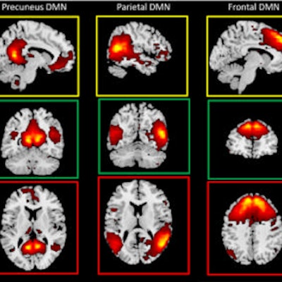
 MR images of the brain's default mode network subcomponents. Images courtesy of RSNA.
In the second study, 20 high school football players with a median age of 16.9 years also wore HITS helmets for one season. Among the subjects, five players had experienced at least one concussion, while the remaining 15 athletes had no such history.
The players underwent an eight-minute magnetoencephalography (MEG) scan before and after the season to record and analyze the magnetic fields produced by brain activity. The researchers then analyzed the MEG power associated with the eight brain regions of the default mode network.
When the five players with a history of concussion were evaluated after the season, the researchers found significantly lower connectivity between default mode network regions. Players with no history of concussion had, on average, an increase in default mode network connectivity.
Based on the results, Davenport and colleagues concluded that concussions from previous years can influence the changes occurring in the brain during the current season. The results also suggest there are longitudinal effects of concussion that affect brain function.
"The brain's default mode network changes differently as a result of previous concussion," Davenport said. "Previous concussion seems to prime the brain for additional changes. Concussion history may be affecting the brain's ability to compensate for subconcussive impacts."
Davenport and Murugesan recommended that longitudinal studies be conducted to follow young football players and to combine MEG and fMRI to better understand the complex factors involved in concussions.