Interventional radiology (IR) offers clinicians and their patients an extremely broad range of minimally invasive image-guided technologies to diagnose, treat, or palliate an ever-expanding number of conditions, many of which previously required more invasive technologies. Interventional radiology can preclude the need for more invasive and costly procedures, help reduce patient recovery time, and provide improved outcomes.
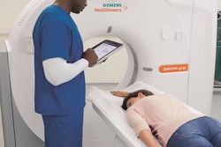
However, minimally invasive procedures performed in imaging departments have increased in complexity. This is, in part, due to the evolution of technology and operating room hybridization, incorporating fixed C-arm tables with image intensifiers, video equipment, and CT or MRI scanners [Martin, 2003]. In addition, patients are often elderly and may come to the interventional radiology suite with complex medical conditions and clinically significant comorbidities.
Many of the procedures performed in the IR suite require procedural sedation and analgesia (PSA). Several agents in different pharmacologic classes are used for sedation and are typically administered intravenously. However, they differ in respect to mechanism of action, potency, onset and duration of action, effect on respiratory drive (central respiratory depression), and individual patient response.
Pharmacologic agents used for procedural sedation and analgesia include anxiolytics, hypnotics, and analgesics. Typical agents administered in this setting are midazolam, propofol, and fentanyl or morphine, respectively. Their use in interventional radiology provides many benefits and is generally considered safe. However, their use can contribute to adverse respiratory events (AREs), which can lead to respiratory compromise due to inadequate patient monitoring or incorrect drug administration/dosage, or as a result of patient-specific variables in the setting of appropriate drug administration/dosage [Martin, 2003; Johnson, 2010].
Studies have shown that certain risk factors predispose patients to periprocedural adverse events. These include opioid naivety, opioid-based analgesic therapy for acute pain management, obstructive sleep apnea, age of at least 65 years, chronic obstructive pulmonary disease, multiple comorbidities, obesity, and deeper levels of sedation and analgesia [Ramachandran, 2011; Taylor, 2015; Fischer, 2013; Khuri, 2011; Aberle, 2017]. Respiratory depression can progress rapidly to failure and arrest [Respiratory Compromise Institute, 2017]. This is a critical event that significantly increases the potential for adverse events, contributes to higher patient costs [Saunders, 2017], and exposes interventional radiologists to litigation [Mavroforou, 2003].
Procedural sedation and analgesia requirements vary considerably depending on patient-specific conditions and the type of procedure. The American Society of Anesthesiologists (ASA) has classified the depth of sedation and analgesia into four stages with specific defining characteristics:
- Minimal sedation (anxiolysis)
- Moderate sedation and analgesia (previously known as "conscious sedation")
- Deep sedation and analgesia
- General anesthesia [Olsen, 2013]
These somewhat arbitrary categories are part of a continuum through which the patient may drift between stages into lighter or deeper states of sedation.
Moderate sedation is often used in the interventional suite, and the number of procedures performed by interventional radiologists using moderate sedation is increasing. By definition, moderate sedation "represents a minimally depressed level of consciousness induced by the administration of pharmacologic agents in which the patient retains a continuous and independent ability to maintain protective reflexes, a patent airway, and the ability to be aroused by physical or verbal stimulation" [American Society of Anesthesiologists Task Force on Sedation and Analgesia by Non-Anesthesiologists, 2002].
This sedation continuum is subject to provider-specific interpretation with no clear demarcation when moderate sedation ends and deeper sedation starts. This can expose a greater risk for adverse events for those not trained in advanced airway management. Accordingly, to prevent deeper levels of sedation, patients require close respiratory monitoring throughout interventional procedures. The ASA's Standards for Basic Anesthetic Monitoring now require capnography for moderate to deep sedation, stating in part that "together with clinical signs ... end-tidal carbon dioxide monitoring ... is effective in detecting hypercapnia or hypercarbia" [American Society for Anesthesiologists, 2011].
In the interventional suite, interventional radiologists are responsible for their patients' overall safety. However, interventional radiologists are understandably focused on the procedure being performed. Therefore, for patients requiring sedation, patient monitoring and the administration of pharmacotherapeutics has become the responsibility of anesthesiologists, certified registered nurse anesthetists, anesthesiology assistants, and registered nurses.
To mitigate some of the risk associated with adverse events, several professional groups, including the ASA (as noted above), the Society of Interventional Radiology, and the Association for Radiologic and Imaging Nursing (ARIN), have issued associational position statements for periprocedural monitoring [American Society for Anesthesiologists, 2011; Baerlocher, 2013; Green, 2016].
Specifically, ARIN endorses routine use of capnography for all patients receiving moderate sedation/analgesia during procedures in the imaging environment because of technical superiority and cost advantages [Green, 2016]. A presedation examination is therefore required for every patient prior to receiving PSA. The presedation evaluation includes a focused history and physical examination to determine the patient's ASA physical status classification score, which is used to identify risk factors that would predispose the patient to AREs [Kohi, 2015].
Current respiratory monitoring with the addition of capnography during sedation procedures is key to detecting respiratory depression before it progresses to respiratory failure. It is important to continually evaluate the adequacy of ventilation and recognize signs of respiratory depression caused by oversedation. Vital signs (blood pressure, pulse, respiratory rate) should also be monitored at prescribed appropriate intervals, together with continuous electrocardiographic monitoring [Kalinowsky, 2005; Tobias, 2011].
Although respiratory monitoring by direct observation alone with pulse oximetry may be effective at detecting adverse events, the interventional setting poses specific challenges. Interventional radiology procedures are often performed in darkened rooms purposefully to allow the proceduralists to view x-ray imaging.
In addition, patients are covered with drapes to maintain sterility and placed in positions that preclude direct visualization of ventilatory function; also, more importantly, staff is often physically separated from the patient during imaging to limit radiation exposure [Long, 2016]. Understandably, patients in IR need to be monitored continuously with pulse oximetry. However, with the inability of pulse oximetry to detect ventilation, the use of capnography is a valuable addition that can assess the patient's ventilatory status by measuring arterial carbon dioxide and detecting real-time respiratory depression and apnea [Brast, 2016].
Continuous pulse oximetry has demonstrated clinical utility for identifying desaturation events and remains the standard of care for monitoring patients who are at risk for adverse respiratory events. However, administration of supplemental oxygen during interventional procedures can maintain arterial oxygen saturation levels close to normal for some time after breathing has stopped and is termed hypoapneic oxygenation [Kalinowski, 2005]. This creates a lag time and results in a pulse oximetry reading that continues to show high saturation rates for several minutes in the presence of hypercarbia [Kohi, 2015]. Therefore, without the visual assessment cues, relying on pulse oximetry alone to monitor ventilatory status may delay the recognition of AREs.
In contrast, capnography provides an immediate picture of a patient's ventilatory status with real-time data that can be used to monitor patients who may be at increased risk for AREs [Ajizian, 2016]. Studies have shown that continuous respiratory monitoring with pulse oximetry and capnography is optimal for the safe administration of opioid analgesia and prevention of adverse events [Overdyk, 2007].
Moreover, respiratory monitoring should be continued into the recovery period, especially when using opioids for postprocedural pain relief [Tuite, 2005]. Because opioids increase the risk of adverse respiratory events in settings where continuous assessment of a patient's ventilatory status is inherently difficult, having the ability to monitor capnography in the interventional suite has the potential to enhance patient safety [Brast, 2016]. Capnography permits an objective measurement of patients' ventilatory status and can identify early evidence of respiratory depression or hypoapneic ventilation, which can promote interventions aimed at preventing an ARE [Long, 2016].
If monitoring identifies an adverse respiratory event, it generally is reversible with prompt intervention [Tobias, 2011]. Management may require a multidisciplinary approach, including administration of supplemental oxygen, airway repositioning maneuvers, drug reversal, and utilization of assisted ventilation when necessary [Chakrabarti, 2006]. Appropriate equipment and drugs required for cardiopulmonary resuscitation and airway support should be available in every IR suite engaging in procedural sedation and anesthesia. In addition, adequate suctioning facilities, airway emergency equipment for pulmonary intubation, and code carts should be available.
Interventional radiology clinicians who administer sedation should also be trained in advanced life support and be competent in using a bag-valve-mask device [Tobias, 2011]. Support can include management of the airway, breathing, circulation, and supplemental oxygen if hypoxemia develops. If necessary, management then proceeds to repositioning the airway or placement of a nasal airway to relieve upper airway obstruction, the application of continuous positive airway pressure to relieve laryngospasm, or bag-valve-mask ventilation for apnea.
If a PSA infusion is running, it should be discontinued when signs of respiratory depression persist. If necessary, additional assistance should be requested as the patient is being stabilized. Rarely, endotracheal intubation and controlled ventilation are needed; however, personnel skilled in such procedures should be readily available. In some instances, reversal opioids and benzodiazepines with naloxone or flumazenil, respectively, may be indicated.
Summary and conclusions
Procedural sedation and analgesia is often required during IR procedures but is associated with an increased risk of adverse respiratory events. AREs can be recognized early by appropriate patient monitoring that should include continuous pulse oximetry and capnography. This allows for timely interventions that potentially can ameliorate adverse events and prevent respiratory compromise.
References
Aberle D, Charles H, Hodak S, O'Neill D, Oklu R, Deipolyi AR. Optimizing care for the obese patient in interventional radiology. Diagn Interv Radiol. 2017;23(2):156-162.
Ajizian S. Capnography outside the OR. Respiratory Care and Sleep Medicine. http://respiratory-care-sleep-medicine.advanceweb.com/Features/Articles/Capnography-Outside-the-OR.aspx. Published April 12, 2016.
American Society of Anesthesiologists. Standards for Basic Anesthetic Monitoring -- Approved by the ASA House of Delegates on October 21, 1986, and last amended on October 20, 2010, with an effective date of July 1, 2011.
American Society of Anesthesiologists Task Force on Sedation and Analgesia by Non-Anesthesiologists. Practice guidelines for sedation and analgesia by non-anesthesiologists. Anesthesiology. 2002;96(4):1004-1017.
Baerlocher MO, Nikolic B, Silberzweig JE, Kinney TB, Kuo MD, Rose SC. Society of Interventional Radiology position statement on recent change to the ASA's moderate sedation standards: capnography. J Vasc Interv Radiol. 2013;24(7):939-940.
Brast S, Bland E, Jones-Hooker C, Long M, Green K. Capnography for the radiology and imaging nurse: a primer. J Radiol Nurs. 2016;35(3):173-190.
Chakrabarti B, Calverley PMA. Management of acute ventilatory failure. Postgrad Med J. 2006;82(969):438-445.
Fischer JP, Shang EK, Butler CE, et al. Validated model for predicting postoperative respiratory failure: analysis of 1706 abdominal wall reconstructions. Plast Reconstr Surg. 2013;132(5):826e-835e.
Green KL, Brast S, Bland E, et al. Association for Radiologic and Imaging Nursing position statement: capnography. J Radiol Nurs. 2016;35(1):63-64.
Johnson S. Sedation and analgesia in the performance of interventional procedures. Semin Intervent Radiol. 2010; 27(4):368-373.
Kalinowski M, Wagner HJ. Sedation and pain management in interventional radiology. Controversies Consens Imaging Interv. http://www.ece.umd.edu/~neil/siddharth/childrens/Sedation_&_Pain_Management_-_GE_Healthcare.pdf. Published 2005.
Kohi MP, Fidelman N, Behr S, et al. Periprocedural patient care. Radiographics. 2015;35(6):1766-1778.
Long M, Green K, Bland E, et al. Capnography monitoring during procedural sedation in radiology and imaging settings: an integrative review. J Radiol Nurs. 2016;35(3):191-197.
Martin ML, Lennox PH. Sedation and analgesia in the interventional radiology department. J Vasc Interv Radiol. 2003;14(9):1119-1128.
Mavroforou A, Giannoukas A, Mavrophoros D, Michalodimitrakis E. Physicians' liability in interventional radiology and endovascular therapy. Eur J Radiol. 2003;46(3):240-243.
Olsen JW, Barger RL, Doshi SK. Moderate sedation: what radiologists need to know. Am J Roentgenol. 2013;201(5):941-946.
Overdyk FJ, Carter R, Maddox RR, Callura J, Herrin AE, Henriquez C. Continuous oximetry/capnometry monitoring reveals frequent desaturation and bradypnea during patient-controlled analgesia. Anesth Analg. 2007;105(2):412-418.
Ramachandran SK, Haider N, Saran KA, et al. Life-threatening critical respiratory events: a retrospective study of postoperative patients found unresponsive during analgesic therapy. J Clin Anesth. 2011;23(3):207-213.
Saunders R, Struys MMRF, Pollock RF, Mestek M, Lightdale JR. Patient safety during procedural sedation using capnography monitoring: a systematic review and meta-analysis. BMJ Open. 2017;7(6):e013402.
Taylor H, Antin N, Grandstrand J, et al. ETCO2 monitoring in high-risk patients in the PACU: a quality improvement project. American Society of PeriAnesthesia Nurses (ASPAN) 34th National Conference. April 26-30, 2015. San Antonio, TX. http://www.aspan.org/Portals/6/docs/ClinicalPractice/2015CSPpdfs/141_Poster.pdf.
Tobias JD, Leder M. Procedural sedation: a review of sedative agents, monitoring, and management of complications. Saudi J Anaesth. 2011;5:395-410.
Tuite C, Rosenberg EJ. Sedation and analgesia in interventional radiology. Semin Intervent Radiol. 2005;22:114-120.
Michael Long works at the department of anesthesiology and critical care medicine at Memorial Sloan Kettering Cancer Center. He graduated from the Nurse Anesthesia Program at Columbia University and completed a Doctorate of Nursing Practice at Barry University. He is an expert in airway management, including intubation, respiratory, and pharmacological support and capnography. He holds a certification in advanced cardiovascular life support and basic life support and has an active certification in the practice of nurse anesthesia in the states of New York and Washington. Long was named one of the 2015-2016 ARIN Stars. He is co-author of ARIN's 2016 position statement on capnography.
Karen Green works as a staff nurse at Metro Vascular Center, a Lifeline vascular access managed center in Philadelphia. She has a bachelor's degree in nursing and a master's in health administration. Green has been active in ARIN for more than a decade, serving in a variety of positions, including president and executive director. She is the recipient of ARIN's Joanna Po Lecture Award and is the lead author of ARIN's 2016 position statement on capnography.
Shawn Brast is the Johns Hopkins Hospital sedation/analgesia subcommittee nursing co-chair and team educator for the Johns Hopkins Lifeline Critical Care Transport Program. Brast has lectured and authored articles on procedural sedation and capnography monitoring. He is co-author of ARIN's 2016 position statement on capnography.