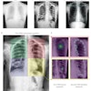
Chinese researchers have found that digital tomosynthesis (DTS) is a better option than conventional digital radiography (DR) for detecting spinal tuberculosis. Tomosynthesis offers better sensitivity than DR, at a lower radiation dose and cost than CT.
Radiologists reading tomosynthesis images rated all images as "excellent" in a group of 55 patients with spinal tuberculosis, according to a study published in the May-June issue of Clinical Imaging, led by Dan Jiao colleagues from China-Japan Union Hospital at Jilin University in Changchun City (Clinical Imaging, Vol. 40:3, pp. 461-464).
The researchers also found that tomosynthesis was better at uncovering a range of pathologies that were signs of spinal tuberculosis, from bone destruction to paravertebral abscesses.
An ancient affliction
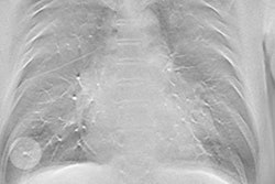
Although spinal tuberculosis is not as well-known as the pulmonary version of the disease, it has afflicted humans for thousands of years, and has even been found in Egyptian mummies, according to a 2011 review by Garg et al in the Journal of Spinal Cord Medicine. It's uncommon in the developed world but can be endemic in some developing nations, particularly in countries with high rates of HIV infection, where pulmonary tuberculosis rates are also high.
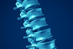
Spinal tuberculosis is particularly common in children and young adults, and it can be debilitating, leading to destruction of the invertebral disk space and collapse of spinal elements, Garg et al reported. If untreated, it can eventually lead to paraplegia, but patients do respond well to treatment with pharmaceutical therapy or surgery.
MRI is typically the best imaging modality for spinal tuberculosis, while CT can demonstrate abnormalities earlier than plain radiography, Garg and colleagues found. But vertebral radiography is often the cornerstone for spinal imaging in many resource-poor countries, they observed.
In the current study, Jiao et al noted that DR is the most common imaging exam for spinal tuberculosis because it's easy to perform and has a low cost profile. DR can visualize many of the telltale signs of spinal tuberculosis, such as narrowing vertebral space and bone destruction.
But DR has low density resolution, and as a 2D modality it can be affected by overlapping tissues and organs. This makes it inadequate for visualizing small lesions and the swelling of soft tissue around the spine and spinal canal.
Digital tomosynthesis, on the other hand, could have advantages because the 3D nature of DTS images could reduce residual blur from out-of-plane structures, the authors wrote.
"DTS images can clearly show the internal structure of complicated parts and the relationship between them and surrounding tissues, and small bone destruction and sequestrum are also shown with the submillimeter plane," they wrote.
Jiao and colleagues decided to test digital tomosynthesis in a group of 55 patients who were seen at China-Japan Union Hospital between 2011 and 2015. They conducted DR and DTS exams on patients before surgery, confirming the presence of tuberculosis with surgical pathology. The patient population included 23 men and 32 women, with a mean age of 40.1 years.
DR exams were conducted with a conventional DR system (Ysio, Siemens Healthcare), while tomosynthesis studies were performed on a DR system outfitted with a tomosynthesis module (SonialVision Safire II, Shimadzu Medical Systems).
After image acquisition, both DR and DTS images were interpreted simultaneously by two radiologists. They evaluated image quality, as well as parameters such as bone destruction, sequestration, narrowing, or disappearing intervertebral space. They also looked for paravertebral abscesses.
The readers rated 83.6% of the DR images as "excellent," but a full 100% of the tomosynthesis images were judged to be excellent. The difference between the two was statistically significant (p = 0.005).
Tomosynthesis also tended to detect more of the anatomical changes that can be caused by spinal tuberculosis, as indicated in the following chart.
| DR vs. DTS for spinal tuberculosis | ||
| Type of pathology | DR | DTS |
| Bone destruction | 85.5% | 100% |
| Sequestration | 32.7% | 69.1% |
| Narrowing or disappearing vertebral space | 90.9% | 90.9% |
| Paravertebral abscess | 36.4% | 63.64% |
In terms of other factors, the researchers found that digital tomosynthesis occupied a middle ground between DR and CT. The image time required for both CT and DTS was about 10 seconds, compared with two seconds for Dr. The cost of DTS was slightly higher than DR, but both were much lower in cost than CT.
With respect to radiation dose, CT unsurprisingly delivered the most dose per exam, at 11.95 mSv, compared with 0.85 mSv for DTS and 0.5 mSv for DR.
The findings indicate that the better image quality and ability to detect pathology inherent in tomosynthesis mean that it could be applied in the diagnosis and follow-up of patients with spinal tuberculosis, serving as a supplementary modality to DR, Jiao and colleagues concluded.
"Compared with DR, the cost and radiation dose of DTS were a little higher, but the image quality and detection rates of lesions were better," they wrote.