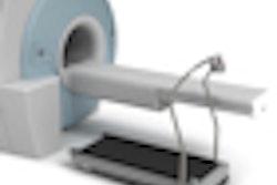
Researchers at Memorial Sloan-Kettering Cancer Center have concluded that a treadmill exercise test before an FDG-PET scan does not increase skeletal muscle FDG uptake and will not adversely affect PET image quality in a clinically significant manner.
The findings contradict the previous belief that physical exercise prior to a whole-body FDG-PET scan would increase plasma glucose insulin levels and deteriorate the subsequent PET image.
"It has been recommended in the past that patients refrain from strenuous physical exercise prior to a PET scan," said lead study author Dr. Ashima Lyall, who presented the research at this month's Society of Nuclear Medicine (SNM) annual meeting. "This has included refraining from treadmill exercise as part of a myocardial perfusion imaging [study] prior to PET."
For patients, the results from the study suggest that it is acceptable to schedule a treadmill stress test on the same day prior to FDG-PET imaging. "This is important in an oncology center like ours, where patients present from all over the country and prefer to have their pretreatment tests scheduled as quickly as possible," Lyall added.
Study population
The retrospective study included 93 patients subdivided into two groups: 48 patients had their PET scans within 12 hours of their cardiac stress test, while 45 patients received their PET scans less than 48 hours after their cardiac stress test, or the next day. The patients had a mean age of 62 years, ranging from 30 to 80 years.
The study also included a third group of 34 control subjects who had no exercise before their FDG-PET scans.
Exercise was continued for an additional two minutes after radiopharmaceutical injection. The duration of exercise for patients in the first two groups was approximately 7.5 minutes, with nine patients (19%) in each group achieving approximately 85% of their maximum heart rate.
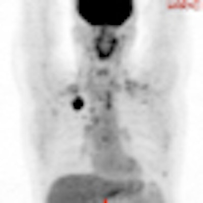
 A 49-year-old male with non-small cell lung cancer received an FDG-PET scan three hours and 45 minutes after a treadmill stress test. The image reveals no conspicuous muscle uptake. SUVmax was 1.2 and SUVmean was 0.7. Image courtesy of Dr. Ashima Lyall.
The mean time difference between the PET scan and the cardiac study was approximately four hours for the same-day group and 38 hours for the next-day group. The mean injection was approximately 15 mCi, with an uptake time of around 17 minutes.
PET scans were performed from the upper thighs to the skull. Researchers calculated standardized uptake values (SUVs) for quadriceps and gluteal muscles in all three groups, as well as for the liver and spine, for background activity. Upper extremity and chest wall muscle uptake was evaluated visually. Myocardial FDG distribution also was compared to SPECT myocardial perfusion imaging findings with a technetium-99m tetrofosmin radiopharmaceutical.
The analysis found only minor, insignificant differences between assessments among the three groups. The only noteworthy exception was SUVmax in the quadriceps muscle in the first group of patients (SUVmax 1.41 ± 0.41, compared with 1.21 ± 0.48 and 1.36 ± 0.22 in the second and third groups, respectively). Image quality and visual assessment of other muscles were deemed similar.
The study did not show any clinically significant increases in muscle uptake for two reasons, according to Lyall.
"One is because there was a relatively short duration and intensity of exercise, although there was enough to provide a myocardial perfusion imaging scan and cause an increase in insulin," she said.
Also, these patients were all in the fasting state, especially the group that was fasting for at least eight to 12 hours. "We think that was partly responsible for the low insulin state," she said.
Only four patients in the first group had at least moderate ischemia, and two patients had infarcts on myocardial perfusion imaging. The researchers found no correlation between myocardial perfusion imaging and myocardial FDG patterns.
Based on the results, Lyall and colleagues concluded that there were small differences in skeletal muscle FDG uptake both in patients with prior treadmill tests and the controls; however, they were not related to the type of exercise or the duration between treadmill exercise and the FDG-PET scans, and they did not appear to be clinically significant.
"There seems to be no clear correlation between the patterns of myocardial FDG uptake and the presence or absence of ischemia on the cardiac test," Lyall said. "Hence, whole-body FDG-PET scans can be performed within a few hours after treadmill test or cardiac stress test without deterioration in PET image quality," and there is no reason to wait for 48 hours between the cardiac stress test and FDG-PET.