Calcifications and other abnormalities of the thoracic aorta on routine chest CT images are highly predictive of cardiovascular events, say the authors of a new study in the November issue of Radiology. The simple model they used to classify multiple aortic findings underscores the value of small abnormalities in uncovering serious disease.
Indeed, the presence of telltale ancillary findings on routine chest CT scans are only the latest reminder, if one were needed, that much serious disease can often be found on CT scans ordered for other purposes. In the case of early heart disease, the availability of so many treatment options makes careful examination of chest CT findings a diagnostic imperative, according to the Dutch authors.
"Several studies have shown that atherosclerotic plaques and calcifications in the thoracic aorta are very prevalent in patients with [cardiovascular disease (CVD)] and stroke in particular," wrote Martijn Gondrie, MD, and colleagues from University Medical Center Utrecht in the Netherlands. "Others have demonstrated that calcification of the descending aorta is related to calcification of the coronary arteries. To date, however, no follow-up studies of investigations of the prognostic value of these aortic abnormalities when they are unexpectedly detected at routine imaging are available" (Radiology, November 2010, Vol. 257:2, pp. 549-559).
An arm of the Prognostic Value of Ancillary Information in Diagnostic Imaging (PROVIDI) study, the goal was to predict CVD by using ancillary aortic findings detected on routine chest CT images to provide a new method of identifying patients who can benefit from timely preventative measures.
A total of 6,975 patients underwent diagnostic contrast-enhanced CT for noncardiovascular indications including rule out of pulmonary embolism, lung cancer or mediastinal disease, and hematologic malignancy. From this group, the researchers examined a representative sample population of 817 patients, in addition to 347 others who experienced a cardiovascular event during a mean follow-up period of 17 months.
CT scans were acquired on a variety of MDCT scanners and were considered acceptable as long as they covered the full length of the thoracic aorta. Slice thickness varied to a maximum of 10 mm. After reviewing the CT scans, readers were asked to assign visual scores for ancillary aortic abnormalities according to the following scales:
- 0 to 8 for calcifications
- 0 to 4 for plaques
- 0 to 4 for irregularities
- 0 to 1 for elongation
The group then constructed four Cox proportional hazard models that included different sum scores for the aortic abnormalities, as well as age, gender, and the CT indication for which the exam was ordered. Finally, they chose the best prediction model and evaluated it externally.
All of the aortic abnormalities had a major prognostic effect, and all of the models performed well (c index range = 0.70-0.72; goodness-of-fit p-value range = 0.45-0.76), they reported. Small findings made a big difference in disease risk.
For example, "with a one-point increase in the aortic calcification score (0-8), the rate of experiencing a cardiovascular event would increase by 25% (hazard ratio = 1.25; 95% confidence interval: 1.17-1.32) throughout the follow-up period, irrespective of the other covariates," the authors wrote. The group chose the simple prediction model based only on calcification score to be the most appropriate for clinical use.
 |
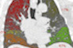
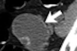
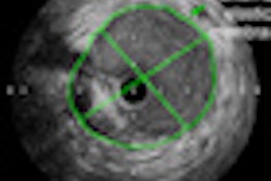
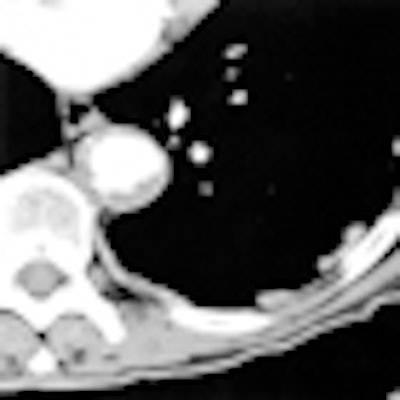
| Aortic findings on transverse CT image shows irregular descending aorta with plaques and some calcified foci. Image republished with permission of the Radiological Society of North America from Radiology, November 2010, doi: 10.1148/radiol.10100054. |
"We chose this model, despite the slightly better statistical fit of the full model, because of its similar c index (0.72 for both models) and goodness-of-fit (p > 0.05 for both models) values and because it is far easier to implement for routine clinical use than is the full model," the group explained. This simple model can be applied to nonenhanced images and involves manual scoring of calcifications only."
Validation of the calcification model in an external dataset also revealed good performance (c index = 0.71; goodness-of-fit p-value = 0.25; sensitivity = 46%; specificity = 76%).
"This simple method ... could be used as an alternative to existing prediction models to identify patients at high risk for CVD in a hospital setting," they wrote.
Subcohort of patients with cardiovascular disease predicted by ancillary aortic findings
|
Individual aortic image characteristics were prognostic for the presence of cardiovascular disease and the occurrence of cardiovascular events. Over the follow-up period, 347 of 817 patients in the subcohort experienced a cardiovascular event. Elongation was present in 7% (n = 59) of patients in the subcohort, and 50% (n = 406) had calcifications in the supra-aortic arteries.
"Individual aortic image characteristics had a prognostic effect in terms of the occurrence of CVD," they wrote. "However, when modeled together, the predictions did not become more accurate," leading to the choice of the simple calcification model.
Reclassification of risk categories could not be considered because the prediction model does not generate recognized threshold values for short-term risks, they added. Sensitivity and specificity were based on the top quartile of predicted risks in the external validation set.
"The sensitivity of 46% (125 of 273 cases) and specificity of 76% (3,669 of 4,815 cases) are only slightly lower than the sensitivity and specificity reported for a more sophisticated CVD risk score for primary care," they noted.
Study limitations included a lack of detailed patient histories or records and the relatively short follow-up time.
"The derived prediction model incorporating ancillary findings of the aorta detected at routine diagnostic CT complements established risk scores and may help to identify patients at high risk for CVD," Gondrie and colleagues concluded. "The timely application of preventative measures may ultimately reduce the number or severity of future CVD events."
By Eric Barnes
AuntMinnie.com staff writer
October 22, 2010
Related Reading
FDG-PET/CT viability scans detect vulnerable aortic plaque, September 27, 2010
CTA screening study shows soft-plaque risk in police, October 4, 2010
Aortic root dilation in athletes requires close surveillance, September 10, 2010
Thoracic and abdominal aortic aneurysms often coexist, July 13, 2010
MRI 'more robust' for assessing aortic root in connective tissue disorders, March 1, 2004
Copyright © 2010 AuntMinnie.com