That conclusion comes from a study led by Dr. Rajesh Gupta, a third-year radiology resident at Stony Brook University Hospital.
Gupta and colleagues conducted a retrospective review of rectal cancer patients at Stony Brook from June 2008 through March 2013. They selected 18 patients (average age, 58 years) who were initially staged with FDG-PET/CT and pelvic MRI within a three-month interval. The results ranged from stage I to III cancer.
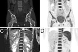
The researchers compared PET/CT and MRI to determine the extent of tumors and local and distant metastases. They then fused PET and pelvic MRI to assess nodal involvement using fusion software.
Five patients (28%) were upstaged based on hypermetabolic lesions seen on PET/CT; three moved from stage III to IV. Four patients (22%) were downstaged because lymph nodes identified on MRI were negative on PET. MRI and FDG-PET/CT examinations were concordant for the remaining nine patients (50%).
MRI provides "excellent anatomical evaluation" of tumor size and extent, while PET/CT offers additional information on metabolic activity, Gupta and colleagues concluded.
"Fusion PET/MRI can provide more accurate staging of local nodal involvement," they wrote in the study abstract. "This may change the initial stage, which can affect treatment options."