Tomosynthesis for general radiography is poised to make a comeback into mainstream diagnostic imaging, its third debut over a 70-year time span. Giving the technique its latest lease on life is the increasing use of flat-panel digital radiography (DR) detectors to acquire a series of images in a matter of seconds, as well as rapid image processing algorithms to reconstruct the images, and PACS diagnostic workstations to display them.
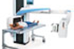
In tomosynthesis, a series of low-dose exposures are made during a single sweep with a fixed or moving detector (depending upon the equipment design) and a tube moving within a limited angular range. The acquired images are processed into slices that show anatomical structures at different depths and angles.
In a single projection radiograph, all the anatomical structures are visible, but there is no sense of depth. In a slice reconstructed via tomosynthesis, only structures within the viewed slice are crisp. The depth and shape of a structure can be determined by viewing multiple slices in sequence in a stack mode.
Most of the attention focused on tomosynthesis recently has been on its use for mammography applications (in which it is still a work-in-progress technology), but vendors and researchers are beginning to recognize its value for general radiography as well.
Tomosynthesis systems are already well-established in Japan, and both GE Healthcare of Chalfont St. Giles, U.K., and Shimadzu Medical Systems North America have digital tomosynthesis equipment that is commercially available, having received 510(k) clearance from the U.S. Food and Drug Administration and the CE Mark in Europe.
GE's version of tomosynthesis is made possible by its VolumeRAD software for the Definium 8000 DR system, introduced in November 2006. Since its release five months ago, more than 100 DR systems with digital tomosynthesis functionality have been sold, including both new Definium 8000 sales and field units that have been modified and upgraded, according to Gerald Schulte, global radiographic marketing manager for GE. The company utilizes an amorphous silicon detector with cesium iodide scintillator on its DR systems.
Shimadzu introduced digital tomosynthesis functionality in Japan in September 2004 with its Sonialvision Safire, a multipurpose digital radiography/fluoroscopy (R/F) system incorporating a selenium-based flat-panel digital detector. The company decided to defer a formal new product introduction in North America until a new model, Sonialvision Safire II, was ready for U.S. distribution this summer, according to Jim Mekker, U.S. tomosynthesis product specialist for Shimadzu Medical Systems North America.
Mekker reports that Shimadzu is now actively selling Safire II, with U.S. installations scheduled for the first quarter of 2008. At the beginning of the 2007 calendar year, 89 Sonialvision Safire I and Safire II systems had been installed in hospitals in Japan, he said.
How it works
The GE system acquires up to 60 individual projection images, and the Shimadzu system collects 75 images in 2.5 seconds at 30 frames per second. The efficiency of digital detectors provides very high-quality images with very low radiation dose, due to high x-ray absorption and low-noise electronics. Compared to a low-dose chest CT (2.5 mSv to 8 mSv), a 10-second lung tomosynthesis study has a dose estimated at 0.77 mSv.
 |
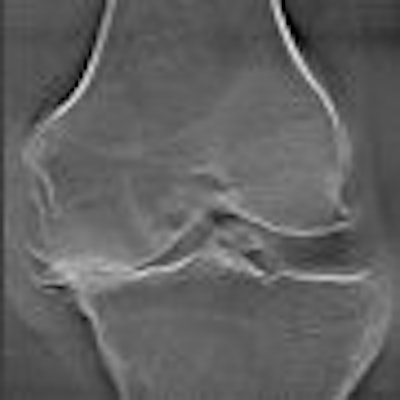
| Single-slice view from a multiple-slice tomosynthesis knee exam. Image courtesy of Shimadzu Medical Systems North America. |
One tomosynthesis pioneer is James Dobbins III, Ph.D., an associate professor of radiology and biomedical engineering at Duke University Medical Center in Durham, NC. Dobbins and colleagues at Duke Advanced Imaging Laboratories patented a tomosynthesis algorithm in the late 1980s, which was never commercially utilized due to the introduction of spiral CT. The introduction of flat-panel digital detectors, with their ability to produce high-quality images, and the potential of rapid reconstruction algorithms renewed interest in the technology.
In 1998, Dobbins and colleagues started to evaluate the feasibility of using digital tomosynthesis for chest imaging. This work led to a research collaboration with GE that contributed to the ultimate commercialization of the VolumeRAD software. Dobbins predicts that with commercial tomosynthesis systems now available, many more clinical studies will begin to be conducted at numerous other healthcare institutions.
Duke is in the final stages of completing the last observer study of a National Institutes of Health grant evaluating the sensitivity and specificity of identifying subtle pulmonary nodules in conventional digital radiographs and tomosynthesis images. Depending upon nodule size, Dobbins estimates that tomosynthesis images will prove to be two to three times more sensitive than conventionally acquired images, and will fit halfway between conventional chest radiography and CT.
"Tomosynthesis is not a replacement for CT," Dobbins noted. "Tomosynthesis is intended to improve chest radiography by enabling radiologists to identify up to 30% more subtle pulmonary nodules. This is important for the millions of people in the world who have a chest x-ray each year."
Duke is planning to perform a cost-effectiveness study in the near future, Dobbins said.
Tomosynthesis in clinical practice
The Princess of Wales Hospital in Bridgend, Wales, U.K., performs chest tomosynthesis after a general chest x-ray has been performed when its radiologists are suspicious of an abnormality.
Dr. Siân Phillips, a consultant radiologist, said that digital tomosynthesis produces superior images for identifying the locations of pulmonary lesions and visualizing their density. It is easier to identify a pleural-based abnormality and differentiate it from a pulmonary abnormality with tomosynthesis, according to Phillips.
"My colleagues and I have also had experience seeing multiple intrapulmonary lesions which cannot be seen on a standard chest radiograph," she said.
 |
| Chest image taken with GE's VolumeRAD tomosynthesis software. Image courtesy of GE Healthcare. |
The radiology department of the Princess of Wales Hospital is also in the planning stage of implementing a clinical trial to evaluate whether digital tomosynthesis alone can diagnose an occult scaphoid fracture without the use of MRI or nuclear medicine. "If we can diagnose a fracture at the initial investigation using a lower-cost procedure, this can have significant economic implications," Phillips said.
Both Phillips and Dr. Ian Scott, one of eight radiologists at Dr. Everett Chalmers Regional Hospital in Fredericton, New Brunswick, Canada, are impressed by the simplicity and speed of acquiring tomosynthesis images. With their system, a radiographer (radiologic technologist) selects the tomosynthesis protocol on the user interface for a chest exam, they noted. Because it takes only an additional 10 seconds to acquire the additional images, the facility's workflow is not affected.
VolumeRAD has 15 anatomical settings that help automate the acquisition process, according to GE's Schulte. These include the chest, abdomen, pelvis, shoulder, hip, knee, hand and wrist, ankle, foot, facial bones, paranasal sinus, temporomandibular, and cervical/lumbar and thoracic spine.
Scott believes that digital tomosynthesis produces exquisite images. "On a day-to-day basis, tomosynthesis is eliminating simple problems and extra procedures that we have routinely had to contend with," he said. "As an example, there is a lot of bony prominence in the areas of the upper anterior sternal margin where the ribs connect to the sternum. Because of this, pseudopulmonary nodules can be projected onto the chest x-ray. If we have any concern about whether we are missing something in the lungs or have concerns about the bony prominence, we'll order a tomo."
Before the DR tomosynthesis equipment was installed in September 2006, the only other choice the radiologists had was to order an apical lordotic x-ray or a CT scan. Scott estimates that approximately 2% of procedures have been averted this way.
There is a high incidence of renal calculi in New Brunswick, and the Dr. Everett Chalmers Regional Hospital operates a shock lithotripsy treatment center that performs kidney stone procedures. The fragments of the kidney stones are routinely tracked until they disappear.
"In conventional imaging, fecal matter in a colon can hide the kidney, and the x-ray procedure may need to be repeated. If we order a tomosynthesis study instead, we are able to 'tomo' through the kidneys and locate where the kidneys and the stones are," Scott said. "This is more efficient for the patient, the radiographer, and the radiologist."
Another tomosynthesis researcher, Michael Flynn, Ph.D., who has led the tomosynthesis research conducted at Henry Ford Hospital in Detroit since 2006, has utilized the technology primarily for orthopedic applications.
Henry Ford Hospital has focused on using digital tomography for musculoskeletal imaging. The radiology department has been particularly progressive in integrating digital radiography into its operations, and had been using flat-panel DR quite extensively, Flynn said. When Mikron Digital Imaging, a regional distributor based in Livonia, MI, made the hospital aware that Shimadzu was seeking a clinical evaluation site in North America, the department thought this would be a good research opportunity.
"We were interested in the fact that this device had a 17 x 17-inch direct selenium flat-plate detector with a 1,000-micron thickness," Flynn said. "We immediately thought of musculoskeletal imaging in the bone detail. We also thought it would be ideal for chest imaging, based on the research from Duke. What I think we didn't know when we first got the unit was whether the exquisite detail in the detector could be fully captured in a tomosynthesis reconstruction. We thought it could be, and have measured this experimentally."
Flynn's primary concern was whether registration of a swinging x-ray tube and a detector moving through a 40° arc could maintain an exact registration. A recorded detail at one angle needs to be back-projected and aligned within one-tenth of a millimeter or finer, otherwise the registration blurs the detail in the reconstruction.
Flynn, as well as Dr. Marnix van Holsbeeck, Henry Ford's director of musculoskeletal radiology and emergency radiology, and Dr. Jamal Ksar, developed protocols for orthopedic imaging. "Initially, we didn't fully appreciate the relationships between the position of a complex structure and both the tomo slices and the direction of the acquisition. As we began our research, we've had to learn how we want to orient the plane relative to the organ and determine the direction we want to scan. This took us three to six months to fully appreciate."
Henry Ford Hospital reconstructs its tomosynthesis images at 1-mm slice increments because Flynn and his colleagues believe that this width shows significant changes in the image. The standard slice thickness is about 3 mm, Flynn explains, but it is weighted toward the center of the 3-mm slice and gets diffusely blurred at the 3/4/5-mm area.
Excelling at small fractures
The early adopters of digital tomosynthesis are convinced it is superior to general radiography and CT in identifying small fractures in the hip, knee, and limbs. One patient, a college basketball player on a highly competitive team, recently presented at the emergency department of Henry Ford Hospital with wrist pain. The x-rays did not show any fracture, but subsequent tomosynthesis images showed a clear crack on the distal radius. Without tomosynthesis, the patient would probably have been diagnosed with a painful sprain and would have continued to play basketball until the crack opened up and the injury worsened.
Scott at Dr. Everett Chalmers Regional Hospital said that tomosynthesis is ordered for simple fractures of the rib, wrist, and tibial plateau fractures of the knee that are suspected but not detected with conventional radiography. The emergency physicians at the hospital are encouraged to order digital tomosynthesis instead of conventional x-rays after hours because of the ease of identifying fractures rather than telephoning the radiologist on call.
"All hairline and simple fractures show up better with tomography," Scott said. "This type of exam usually requires less movement by a patient, who may be in considerable pain." The images still may contain artifacts, such as a tiger-stripe artifact that appears with images of bone fractures, he noted.
Other applications being evaluated include postmyelogram spine studies used in lieu of CT at Henry Ford Hospital, and digital tomosynthesis instead of MRI of the shoulder and shoulder arthrograms at Chalmers Hospital.
Now that commercial systems are available, Dobbins believes that radiologists will begin to use digital tomosynthesis for chest and musculoskeletal imaging on a regular basis. He thinks that it is just a matter of time for widespread clinical validation to support adoption of the technology.
As to whether digital tomosynthesis with CAD can become an affordable screening tool for lung cancer, Dobbins suggests that this is premature. The National Lung Screening Trial will need to be completed before it is determined if this resurrected technology can become part of a major public health screening program. However, its future as an adjunct to conventional radiography seems bright.
By Cynthia Keen
AuntMinnie.com contributing writer
October 29, 2007
Related Reading
Duke researchers combine DR tomosynthesis with 3D CAD, June 4, 2007
Dual-energy digital x-ray still looking for acceptance, February 22, 2007
Copyright © 2007 AuntMinnie.com