Researchers at New York University's (NYU) School of Medicine have found that the combination of functional information from F-18 FDG-PET and anatomic localization from MR imaging can provide a valuable diagnostic method for the detection of primary and recurrent breast cancer.
The study noted that fused PET and MRI images substantially increased the specificity of MRI, but, at the same time, decreased MRI's sensitivity in the group of patients analyzed in the NYU research. Results were recently published by the New York City-based authors in the Journal of Nuclear Medicine.
While MRI has shown high sensitivity in the detection of breast cancer, the modality has not performed as well on its own regarding specificity. To boost MRI's specificity, the NYU group sought to determine how well the addition of F-18 FDG-PET's metabolic information would improve breast lesion characterization.
Increased specificity of MRI, the study noted, could also potentially spare a patient from more invasive procedures, such as fine-needle aspiration, biopsy, or resection, as well as avoid follow-up examinations.
As part of the research, the NYU group also designed a prototype device, which would allow a patient to be PET imaged in the prone position as the MRI scans. A patient's breasts would hang down freely simply by way of gravity.
Breast MRI examinations were performed with the patient in the prone position, using either a 1.5- or 3-tesla MRI system (with a dedicated surface breast coil) from Siemens Medical Solutions of Malvern, PA.
Protocol and methods
The protocol employed an imaging sequence that included a sagittal T1-weighted localizing sequence followed by a sagittal T2-weighted sequence. Next, a T1-weighted 3D, fat-suppressed fast spoiled-gradient-echo sequence was performed before and four times after a rapid bolus injection of 0.1 mmol per kilogram body weight of gadolinium dimeglumine at an injection rate of 2.0 mL/s.
Image acquisition for the study began immediately after administration of the contrast and saline bolus. Total examination time per breast was approximately 15 minutes.
Additional images were acquired using a Siemens PET/CT system, which featured a high-resolution lutetium oxyorthosilicate crystal PET scanner integrated with a six-detector CT scanner. Scanning was started 45 minutes after injection. No intravenous contrast agent was administered.
For prone patient imaging, the positioning device allowed the same patient geometry as the breast MRI coil to provide identical breast image configuration for both PET and MRI scans.
The report also noted that "when no lesions were seen on CT or no abnormal F-18 FDG uptake was seen on PET images, then ROIs (regions of interest) were placed in the region of the abnormalities indicated by the MRI. Their location was determined by using the MRI to approximate the location on the coregistered CT scan."(JNM, April 2007, Vol. 48:4. pp: 528-537).
 |
| A 44-year-old woman shown in 3D with arrow indicating 2-cm lesion confirmed as moderately differentiated IDC. Yellow, shown transparent, is MRI scan with fused PET lesion and other high-activity regions (liver and spine) superimposed in red. Lesion is inside left breast, whereas liver and spine are in front of and beneath MRI scan. Image (A) shows a 3D view from one angle with level of PET activity set at one particular level. Image (B) shows a 3D view from a different angle with level of PET activity set at a higher level than in A. |
Study analysis
In all, 22 patients were evaluated from January 2005 and January 2006. Twenty-nine breasts with a total of 45 suspicious lesions detected through MRI scans were included in the analysis. Cancer was confirmed in 22 breasts, while no cancer was detected in the other seven breasts.
Seven of the 45 lesions were seen in the contralateral breast. The lesions ranged in size from 0.6 to 10 cm, with a mean of 2.5 cm.
NYU researchers reported that mastectomy was performed on 13 breasts; lumpectomy or surgical excision was used on five breasts; fine-needle aspiration on two breasts; and core biopsy in the one remaining case. For the other eight breasts, the study noted, clinical and radiologic follow-up was performed for at least one year on 11 lesions.
In the study's analysis of the 45 lesions, sensitivity and specificity of MRI were 92% and 52%, respectively, while sensitivity and specificity for fused MRI and PET were 63% and 95%, respectively.
The positive predictive value (PPV) for MRI alone was 69%, while the negative predictive value (NPV) was 85%. For fused MRI and PET, the PPV was 94% and NPV was 69%. Both PET and MRI were able to detect increased metabolic activity in lesions.
 |
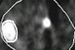
| A 27-year-old woman, presented with one enhancing mass in each breast. Axial (top), coronal (middle), and sagittal (bottom) slices are shown at level of a 2.2-cm mass detected in left breast (arrows) on both MRI and PET scans and found to be poorly differentiated DCIS at mastectomy. Image (A) is an unregistered PET scan. Image (B) is the original MRI scan. Image (C) shows registered PET scan superimposed on original MRI scan. As can be seen in (A) on axial and coronal views, mass in right breast was correctly not seen on PET scan as determined also by mastectomy. Reprinted with permission from "Improving Specificity of Breast MRI Using Prone PET and Fused MRI and PET 3D Volume Datasets," Linda Moy, Fabio Ponzo, Marilyn E. Noz, Gerald Q. Maguire Jr., Antoinette D. Murphy-Walcott, Abby E. Deans, Mary T. Kitazono, Laura Travascio and Elissa L. Kramer, April 2007, Journal of Nuclear Medicine. |
As for the role of PET in the evaluation of suspicious lesions, 14 of 45 lesions were categorized as known cancer. The mean size of the lesions was 2.7 cm, ranging in size from 1.4 cm to 5.2 cm.
The NYU study stated that its preliminary results "suggest that prone PET breast imaging combined with a fusion technique facilitates a better correlation of lesions." One red flag raised by the report was the trend that while specificity increased, sensitivity diminished.
Research limitations
The study cited some limitations, such as PET not showing increased activity at sites of ductal carcinoma in situ (DCIS) on a consistent basis. PET, however, identified all of the cancers that were intraductal carcinoma as hypermetabolic.
The report recommended more research be conducted to determine the potential of PET in both DCIS and invasive lobular carcinoma.
In addition, the study found that the "biggest limitation in comparing prone MR images with supine PET images is the loss of normal landmarks. Marked differences in patient positioning result in unreliability of the usual landmarks -- the quadrant of the breast where an abnormality is located and the distance of the lesion from the nipple."
While fusing PET scans and MR images may increase the specificity of breast MRI, images must be take in the same prone position. The study concluded that the breasts "become markedly distorted when supine/prone images are registered and it is challenging to identify the corresponding lesion in all three orthogonal planes of the fused images."
By Wayne ForrestAuntMinnie.com staff writer
August 17, 2007
Related Reading
Presence of invasive cancer may cause MR-guided biopsy to miss DCIS, August 16, 2007
MRI beats US for breast screening of at-risk women, but yields more biopsies, August 6, 2007
MRI becoming more efficient in breast cancer detection, May 18, 2007
MRI useful for detecting cancer in contralateral breast, March 28, 2007
Dual-time-point breast PET increases diagnostic accuracy, December 2, 2005
Copyright © 2007 AuntMinnie.com