Diaphragmatic Rupture / Traumatic Herniation of the Stomach:
Clinical:
Penetrating trauma is the most common cause of diaphragmatic injury.
Only 3% to 6% of
blunt trauma patients have diaphragmatic ruptures, and these patients
frequently have
multiple other injuries (44-100%) [6,9,11]. Blunt diaphragmatic rupture
remains undiagnosed at initial presentation in 7-66% of cases
(particularly right sided rupture) [16]. The injury is the result of a
sudden increase in intraabdominal or intrathoracic pressure against a
fixed diaphragm [15]. Other postulated mechanisms are a shearing effect
associated with lateral impacts [16]. The left hemidiaphragm (77-90% of
cases [8]) is affected
more than the right, probably as a result of a protective effect from
the liver. and an area of congenital embryologic weakness in the
posterolateral left hemidiaphragm [16]. Most tears from blunt trauma
are longer than 10 cm (whereas penetrating injuries tend to create
short tears (1-2 cm) [16].
Most
diaphragmatic ruptures originate in the posterolateral portion of the
diaphragm at the
junction of its tendon and the posterior leaves. The predilection for
tearing to occur at
this anatomic site is attributable to the congenital weakness of the
fusion between the
costal and lumbar muscular attachments of the diaphragm [1]. Blunt
diaphragmatic rupture occurs with approximately equal frequency and
injury pattern in adults and children [16].
Viseral herniation occurs in up to 95% of left sided lacerations, but may be delayed, and initial signs may go undetected or not become apparent for months or years. The stomach is the most frequent abdominal viscus to herniate [8,16]. Roughly three-quarters of patients with diaphragmatic rupture have other intra-abdominal injuries (splenic injury in 60%), and 42% have pelvic fractures [8,12]. Nearly 70% of the lesions are initially missed, possibly secondary to overlying pulmonary (contusion or atelectasis) and pleural (effusion) abnormalities. Mechanical ventilation in these patients also helps to expand the lungs which may impede herniation of the abdominal contents. [2,3,10] Unrecognized diaphragmatic rupture can be complicated by bowel strangulation or obstruction and this is associated with high morbidity and mortality [13,14]. Among patients with blunt diaphragmatic rupture, the overall mortality resulting from other life-threatening injuries varies from 12-42% [16]. Treatment is surgical repair [14].
X-ray:
View cases of diaphragmatic rupture
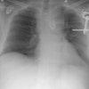
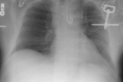
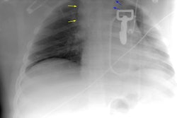
CXR: Up to 77% of patients with diaphragmatic rupture will have an abnormal CXR, however, the diagnosis is suspected in less than 50% of the cases on the basis of the radiographic findings [7]. CXR's have a sensitivity of 45% for left sided rupture and only 17% for right sided rupture [14]. Specific signs of left diaphragmatic rupture include a intrathoracic herniation of a hollow viscus and an abnormal course of a nasogastric tube above the hemidiaphragm [12]. Other signs which suggest a diaphragmatic injury include a left pneumothorax, a basal pulmonary opacity, slight irregularity to or loss of the diaphragmatic contour, shift of the mediastinum to the opposite side, and an elevated left hemidiaphragm. With penetrating injury, a bullet near the diaphragm or an entrance wound with a bullet or a blade that indicates a path through the diaphragm should also raise the suspicion of diaphragmatic tear. A barium study can be performed to confirm the presence of bowel loops in the thoracic cavity.
Computed tomography: Standard CT is limited in the diagnosis of diaphragmatic rupture due to the transaxial nature of the images (Sensitivity 61-73% [6]). The limitations of standard CT for the diagnosis of diaphragmatic rupture were primarily due to difficulty in differentiating abdominal organs beneath an elevated hemidiaphragm from visceral or omental herniation through a post-traumatic defect [7].
Helical CT has been reported to have a sensitivity of 71% to 87%, specificity of 75% to 100%, positive predictive value of 81%, a negative predictive value of 81%, and an accuracy of 83% [11,12,16]. Sensitivity is better for left side rupture (78%) versus right sided injury (50%) [9]. Multidetector CT with multiplanar sagittal and coronal reformatted images has been shown to increase the sensitivity to 71- 92%, and specificity to 98-100% [7,16], although other authors do not feel that these images aid in exam interpretation [11]. Reformatted images may very useful in identifying liver herniation [9]. CT can still be falsely negative in patients that have tears without associated intrathoracic herniation of abdominal contents [9]. CT findings of diaphragmatic tear include:
1- Discontinuity of the diaphragmatic outline (most common finding-
73-82% of cases
[8]). However, the diagnosis should not be exclusively based on this CT
finding
[12]. Discontinuity of the diaphragm can be found in 6% of the general
population [14]. In elderly patients the diaphragm may be discontinuous
in up to 35% of normal
patients, and therefore, this sign may not be as reliable in the
elderly [3].
Women and patients with underlying emphysema also have a higher
incidence of normal discontinuity [14]. Note that non-pathologic
diaphragmatic discontinuity is generally not seen in patients under the
age
of 39 years [7].An upper hemidiaphram contour that rises more than
4-6cm above the level of the contralateral hemidiaphragm is also
suggestive of diaphragmatic rupture [16].
2- Bowel loops or omental fat in the thoracic cavity. The stomach
and colon
are the most common visera to herniate on the left side [12]. An
abnormal U-shaped course of a nasogastric tube with the tip projecting
above the level of the diaphragm is a clue to intrathoracic gastric
herniation [16].
3- "Collar" sign- Viscera have a waist-like constriction at the site of herniation and this is referred to as a "collar" sign [9]. On the right side, the collar sign can appear as a focal indentation of the liver [12]. The collar sign is 67% sensitive for left-sided rupture and 50% sensitive for right sided rupture when sagittal and coronal reconstruction images are obtained [14]. The "sandwich" sign refers to contrast filled stomach which has herniated into the chest and folded upon itself.
4- "Dependent viscera" sign- The bowel or viscera are positioned against the posterior ribs with obliteration of the costophrenic recess (herniated abdominal organs are in direct contact with the posterior chest wall without interposition of lung) [14,16]. This occurs due to the absence of the posterior support of the diaphragm which allows the viscera to "fall" against the posterior ribs [11]. The sign is 100% sensitive for the diagnosis of diaphragmatic rupture [14], but other authors quote a sensitivity of 64-100% for left sided rupture and a specificity of 98-100% [16].
5- Thickening of the diaphragm or diaphragmatic crus [11,13]
6- Hump and band sign- the hump and band sign result from herniation
of the liver through a right sided defect [16]. The hump sign is a
variation of the left sided collar sign and refers to the shape of the
herniated liver above the level of the diaphragm [16]. The band sign
corresponds to a linear area of hypoattenuation that transects the
herniated liver between the torn edges of the diaphragm [16]. The hump
and band signs are best demonstrated on coronal and sagittal
reconstruction images [16]. Reported sensitivites are 50-83% for the
hump sign and 33-42% for the band sign [16]. This sign should not be
confused with a diaphragmatic eventration- a condition that most often
affects the left hemidiaphragm- in which the diaphragm is thinned, but
remains intact [16].
Magnetic resonance imaging: MR imaging may have certain advantages over CT due to it's ability to image in the coronal and sagital planes. The hyperintense abdominal and mediastinal fat permit optimal visualization of the hypointense diaphragm on T1 sequences. Experience, however, is limited [4].
REFERENCES:
(1) Arch Surg 1963; 86: 989-994
(2) J Thorac Imag 1995, 10: p.150-152
(3) Semin Ultrasound CT MR 1996 Apr;17(2):114-118
(5) RadioGraphics 1998; 18: 49-59
(6) AJR 1998; Aoki AA, et al. Traumatic rupture of the right hemidiaphragm in an automobile accident victim. 171: 386 (No abstract available.)
(7) Radiographics 1998; Van Hise ML, et al. CT in blunt chest trauma: Indications and limitations. 18: 1071-1084
(8) Radiographics 1998; Kuhlman JE, et al. Radiographic and CT findings of blunt chest trauma: Aortic injuries and looking beyond them. 18: 1085-1106
(9) AJR 1999; Killeen KL, et al. Helical CT of diaphragmatic rupture caused by blunt trauma. 173: 1611-1616
(10) AJR 2001; Carter YM, et al. Delayed recognition of diaphragmatic rupture in a patient receiving mechanical ventilation. 176: 428
(11) AJR 2002; Larici AR, et al. Helical CT with sagittal and coronal reconstructions: accuracy for detection of diaphragmatic injury. 179: 451-457
(12) Radiographics 2002; Iochum S et al. Imaging of diaphragmatic injury: a diagnositic challenge. 22: S103-S118
(13) AJR 2005; Nchimi A, et al. Helical CT of blunt diaphragmatic rupture. 184: 24-30
(14) Radiology 2006; Cantwell CP. The dependent viscera sign. 238: 752-753
(15) Radiographics 2008; Kaewlai R, et al. Multidetector CT of blunt
thoracic trauma. 28: 1555-1570
(16) Radiographics 2012; Desir A, Ghaye B. CT of blunt diaphragmatic
rupture. 32: 477-498