Epstein-Barr-virus-associated lymphoproliferative disease of the lung: CT and histologic findings.
Collins J, Muller NL, Leung AN, McGuinness G, Mergo PJ, Flint JD, Warner
TF, Poirier C, Theodore J, Zander
D, Yee HT
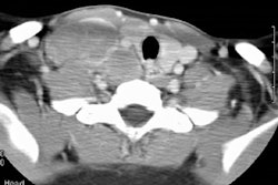
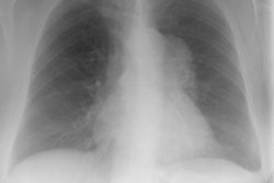
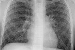
PURPOSE: To assess the computed tomographic (CT) and histologic findings of intrathoracic lymphoproliferative disease (LPD) associated with the Epstein-Barr virus (EBV). MATERIALS AND METHODS: The authors retrospectively reviewed the CT scans of the chest and the pathologic specimens obtained in 24 patients with histologically proved intrathoracic LPD and with positive serologic findings or immunohistochemical staining for EBV. Five patients had acquired immunodeficiency syndrome (AIDS); one had common variable immune deficiency; and 18 were receiving immunosuppressive therapy for heart, lung, or heart-lung (n =15) or bone marrow (n = 2) transplantation and vasculitis (n = 1). RESULTS: Final diagnoses included malignant lymphoma (n = 15), polyclonal LPD (n = 8), and hyperplasia of bronchus-associated lymphoid tissue (n = 1). CT findings included multiple nodules (n = 21), lymphadenopathy (n = 9), areas of groundglass opacification (n = 8), septal thickening (n = 7), consolidation (n = 5), pleural effusion (n = 4), and solitary endobronchial lesion (n = 2). The nodules were 2-4 cm in diameter, involved mainly the middle and lower lung zones, and frequently had a predominantly peribronchovascular (n = 15) or subpleural (n = 14) distribution. CONCLUSION: EBV-associated LPD may range from benign lymphoid hyperplasia to high-grade lymphoma. The most common CT manifestation consists of multiple nodules, frequently in a predominantly peribronchovascular or subpleural distribution.