Radiographics 1997 Jan;17(1):47-58. Interpretation of chest radiographs in AIDS patients: usefulness of CD4 lymphocyte counts.
Published erratum appears in Radiographics 1997 May-Jun;17(3):804
Shah RM, Kaji AV, Ostrum BJ, Friedman AC
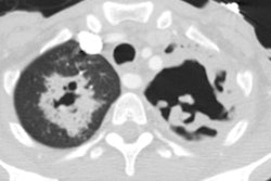
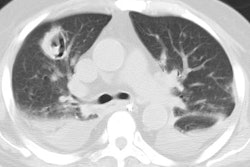
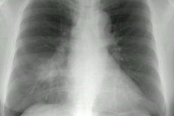
Specific infections and neoplasms that are complications of acquired immunodeficiency syndrome (AIDS) occur within various CD4 lymphocyte count ranges. Knowledge of how these counts correlate with radiographic appearances of these entities can limit the differential diagnosis because certain conditions are uncommon above a specific count. In patients with CD4 lymphocyte counts above 200 cells/mm3 and radiographic findings of cavitary and noncavitary consolidation, bacterial pneumonia and Mycobacterium tuberculosis are the major diagnostic considerations. As the CD4 lymphocyte count falls, these infections are still common; however, cavitation is seen less frequently with Mycobacterium tuberculosis, and unusual bacterial infections, including those caused by Rhodococcus equi and Nocardia asteroides, should be considered. In patients with counts below 200 cells/mm3, Pneumocystis carinii pneumonia is the most common infection, usually manifesting radiographically as a reticular interstitial pattern. At CD4 lymphocyte counts of 50-200 cells/mm3, disseminated fungal infection and Kaposi sarcoma become prevalent. In patients with advanced AIDS and counts below 50 cells/mm3, radiographic nodular or reticular patterns may indicate AIDS-related lymphoma and cytomegalovirus and Mycobacterium avium-intracellulare infections. When CD4 lymphocyte counts are applied to interpretation of chest radiographs in AIDS patients, the working differential diagnosis of a radiographic pattern can be tailored to the clinical situation of a given patient.
PMID: 9017798, MUID: 97170269

