Congenital > Sequestration

Bronchopulmonary Sequestration:

View cases of bronchopulmonary sequestration

Clinical:

A pulmonary sequestration represents isolated lung tissue which lacks a normal communication to the tracheobronchial tree, but contains alveoli and bronchi. The lesion probably arises from an accessory foregut lung bud which has the ability to induce the primitive mesenchyme to form normal pulmonary tissue [12]. If accessory the lung bud persists, the sequestration may communicate with the foregut- especially the esophagus, and less commonly the stomach or biliary tree. The most common location for a sequestration is the posterior basilar segment of the lower lobes (L > R); they are rarely upper lobe. The arterial supply to a pulmonary sequestration is via the SYSTEMIC circulation (usually from a branch arising from the descending of aorta). There are 2 types of sequestrations:

1- Intralobar (ILS): ILS is more common than extralobar sequestration and accounts for 75% of cases. An intralobar sequestration is embedded within normal lung (within the visceral pleura) and has no separate pleural covering. Associated congenital anomalies are seen less commonly than in patients with ELS and are reported in 6-12% of patients [10]. The lesion occurs almost equally in adults and children. It is rarely described in infants or neonates. Although it may be a congenital lesion, its lack of association with other congenital anomalies, the presence of chronic inflammation on histologic analysis, and its uncommon occurrence in infants all suggest ILS to be an acquired lesion. Patients typically have a history of recurrent pulmonary infection and chronic inflammation and fibrosis are often seen on histologic analysis. It is postulated that persistent bronchial obstruction leads to secondary infection and the chronic inflammation eventually leads to disruption of the normal pulmonary arterial supply. Pleuritis and increased vascularity result in parasitization of a systemic arterial supply via normal occurring aortic branches to the visceral pleura in the pulmonary ligament. Its blood supply drains via the pulmonary veins to the left atrium (95% of cases) which results in a unique left-to-left shunt [9]. The lesion may have a fistulous communication with the tracheobronchial tree. About 98% of cases occur in the lower lungs and there is a slight left sided predominance (56-64%). Treatment is surgical resection in order to avoid complications such as recurrent infection or hemorrhage. [1]

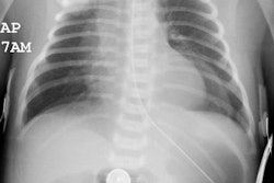
2- Extralobar (ELS): Extralobar sequestrations account for about 25% of cases of sequestration [7]. ELS is a congenital mass of non-functioning pulmonary tissue that has no normal connection to the bronchial tree or pulmonary arteries. The lesion is surrounded by its own pleural covering (which prevents collateral drift) [10]. The lesion usually presents in infants and children (60% of cases by age 6 months). Males are affected more than females (4:1) [8]. Patients with extralobar sequestrations are generally asymptomatic, but come to medical attention as a result of associated congenital anomalies which are found in over 50% of cases (such as diaphragmatic hernia, congenital heart disease, and CCAM type II) [10]. In the absence of other anomalies, the sequestration frequently becomes superinfected and most patients present by age 10y. In the newborn the lesion may produce respiratory distress or CHF secondary to shunting. Other symptoms include dyspnea, cyanosis, and feeding difficulties in a newborn due to mass effect. Older patients may complain of pain, hemoptysis, or superinfection. ELS usually drains via the azygous system to systemic veins and subsequently the right atrium (over 80% of cases) producing a left-to-right shunt [10]. The remaining cases drain via the pulmonary circulation to the left atrium [10]. Extralobar sequestrations have also been found within or below the diaphragm (5%), in the retroperitoneum (2-5%) (4), mediastinum, and intrapericardial. Associated anomalies are common (65% of cases [9]) and include: Ipsilateral diaphragmatic hernia (found in 30% of cases) or a bronchogenic duplication cyst (which is typically interposed between the bronchi and the sequestration). Cardiovascular and renal anomalies can also be seen. Treatment is surgical resection or coil embolization [6].

X-ray:

Prenatal Ultrasound: Extralobar sequestrations can be seen as early as 16 weeks gestation [8]. They typically appear as a well defined triangular echogenic mass [8]. An ipsilateral pleural effusion is seen in 6-10% of cases [8]. Cysts can be seen, particularly in hybrid lesions that combine elements of sequestration and congenital cystic adenomatoid malformation [8]. Both types of BPS may be associated with polyhydramnios and in utero hydrops fetalis due to compression of the esophagus and thoracic venous structures if the lesion is large [11]. However, the lesion may also involute during gestation [11].

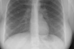
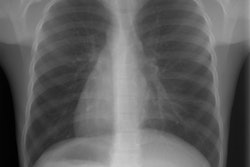
CXR: On plain film an ELS typically appears as a solid mass with a round or pyramidal shape. It is usually between the diaphragm and the lower lobes and is most commonly left sided (60-90%). It can appear as a homogeneous mass or a small bump on the posterior medial hemidiaphragm [7]. The lesion may be subtle on CXR [7]. About 10% will have an ipsilateral pleural effusion. On US an extralobar sequestration appears as a homogeneous, echogenic mass. Scanning may be able to demonstrate a systemic feeding vessel. An UGI exam will exclude communication with the GI tract. Angiography will demonstrate an abnormal systemic arterial supply (most commonly arising from the aorta).

On plain film an ILS appears as a radiographic density/consolidation, soft tissue mass, or cystic/multicystic abnormality within the lower lung. An air-fluid level(s) can be identified within the lesion in 25-33% of cases. Bronchography, although rarely performed today, demonstrates no normal connection to the tracheobronchial tree. Normal bronchi will be displaced about the lesion.

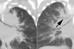
Computed tomography: ELS usually appear as a well-defined, solid, retrocardiac mass in the posterior cardiophrenic or costophrenic angle [10]. Air bronchograms are usually absent with ELS [10]. ILS tend to be more heterogeneous and less well defined often as a complex, single or multicystic mass. The cysts often contain air-fluid levels. Focal bronchiectasis, areas of atelectasis, and cavitation may also be seen within the lesion [10]. Adjacent emphysematous changes are common (the areas can be aerated via collateral drift, but lack of normal bronchial communication results in air-trapping with exhalation) [7]. Calcification of either type of sequestration is unusual [10].

CT, MRI, and angiography can all be used to demonstrate the systemic arterial supply to the lesion most commonly arising from the lower thoracic (75%- located within the leaves of the inferior pulmonary ligament) or upper abdominal aorta/celiac axis (20%). Multiple feeding vessels are found in 16% of cases. Venous drainage occurs through the pulmonary veins of the involved lobe into the left atrium in 95% of cases. In 5% of cases, systemic venous drainage occurs. When predominantly cystic differential considerations include pulmonary abscess, bronchiectasis, bronchogenic cyst, congenital cystic adenomatoid malformation.

Magnetic resonance imaging: On prenatal MR, the lesions usually appears as a solid, well-defined, uniform, hyperintense mass on T2-weighted images [11].

MR does not demonstrate the lesion as clearly as CT, however, 3D breath-hold contrast enhanced MR angiography can offer excellent evaluation of the aberrant arterial supply to the lesion [7]. On prenatal MR, the normal fetal lungs are homogeneous and have a high signal on T2 images (because they are filled with amniotic fluid) [8]. A sequestration typically appears as a well defined mass in the chest that has a T2 signal higher than that of normal lung, but lower than that of free amniotic fluid [8]. 

REFERENCES:

(1) Radiographics 1997; 17: 725-745

(2) AJR 1990; Imaging of pulmonary sequestration. 154(2), 241-249 (No abstract available)

(3) Radiology 1990; 176:375-79

(4) AJR 1997; 168: 1277-81

(5) Radiographics 1999; Ceola AF, et al. US case of the day. 19: 817-819

(6) AJR 2000; Tokel K, et al. Coil embolization of pulmonary sequestration in two infants: A safe alternative to surgery. 175: 993-995

(7) AJR 2000; Ko SF, et al. Noninvasive imaging of bronchopulmonary sequestration. 175: 1005-1012

(8) AJR 2003; Dhingsa R, et al. Prenatal sonography and MR imaging of pulmonary sequestration. 180: 433-437

(9) Radiographics 2003; Konen E, et al. Congenital pulmonary venolobar syndrome: spectrum of helical CT findings with emphysis on computerized reformatting. 23: 1175-1184

(10) Radiol Clin N Am 2005; Paterson A. Imaging evaluation of congenital lung abnormalities in infants and children. 43: 303-323

(11) Radiographics 2010; Daltro P, et al. Congenital chest malformations: a multimodality approach with emphasis on fetal MR imaging. 30: 385-395

(12) Radiographics 2010; Biyyam DR, et al. Congenital lung abnormalities: embryologic features, prenatal diagnosis, and post natal radiologic-pathologic correlation. 30: 1721-1738

Latest in Congenital
Page 1 of 9
Next Page