Systemic sclerosis: using high-resolution CT to detect lung disease in children.
Seely JM, Jones LT, Wallace C, Sherry D, Effmann EL
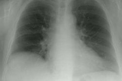
OBJECTIVE: The purpose of this study was to determine the prevalence of interstitial lung disease and the severity of disease in children with systemic sclerosis using high-resolution CT (HRCT). SUBJECTS AND METHODS: Eleven children (mean age, 11 years) with scleroderma underwent HRCT, chest radiography, and pulmonary function testing. Eight of these 11 patients also underwent follow-up HRCT. HRCT studies were assessed by two observers for ground-glass attenuation, honeycombing, and other abnormalities. Profusion scores for ground-glass attenuation and honeycombing were determined by multiplying severity of disease by the percentage of lung involvement. RESULTS: Chest radiographs predicted interstitial lung disease in only two patients, whereas HRCT showed interstitial lung disease in eight patients (p = .05). On HRCT, ground-glass attenuation was found in eight patients (73%), honeycombing in five patients (45%), linear opacities in six patients (55%), and subpleural micronodules in seven patients (64%). By the end of the study, 10 patients (91%) had evidence of interstitial lung disease on HRCT. Overall, profusion scores for these 10 patients showed four patients with mild, one with moderate, and five with severe disease. Also, HRCT revealed worsening disease in three of eight patients. We found no correlation between duration of scleroderma and severity of interstitial lung disease (p > .02). Seven patients with evidence of lung disease on HRCT had abnormal results on pulmonary function tests; patients with the highest scores for ground-glass attenuation had the most abnormal results on pulmonary function tests (p < .01). CONCLUSION: HRCT shows significant pulmonary disease in children with systemic sclerosis, revealing abnormalities in 91% of our patients. Pulmonary disease should be suspected in children with scleroderma, even if the chest radiograph has normal findings.