Microscopic Polyangiitis:
Clinical:
Microscopic polyangiitis (MP) is an ANCA-associated
nongranulomatous necrotizing small vessel systemic vasculitis that
lacks an eosinophilic component [1,2,3]. It is the most common
cause of pulmonary-renal syndrome (diffuse pulmonary hemorrhage
and glomerulonephritis) [1,2,3]. The disorder is primarily renal,
and less commonly pulmonary [1]. The median age of onset is 50
years [2]. There is typically a long prodromal phase of symptoms
such as fever and weight loss [2,3]. More than 90% of patients
then have a rapidly progressive glomerulonephritis at presentation
[1]. Pulmonary involvement occurs in approximately 25-50% of
patients [2]. Diffuse pulmonary hemorrhage occurs in 10-30% of
patients and is commonly present at time of presentation
[1]. Pulmonary symptoms include hemoptysis and shortness of
breath [1]. Other manifestations include skin lesions, peripheral
neuritis, and GI hemorrhage [1]. The musculoskeletal system,
heart, and eyes can also be involved [3]. On lab analysis 40-80%
of patients are p-ANCA positive [anti-MPO] (sensitivity 35-70%)
[1,2].
Histolgically, a necrotizing vasculitis most often affects venules, arterioles, and capillaries [3]. Pulmonary capillaritis manifests as interstitial neutrophilic infiltration and causes necrosis of the alveolar and capillary walls which results in diffuse alveolar hemorrhage [3].
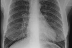
X-ray:
CXR: The radiologic features reflect DAH with patchy or bilateral diffuse airspace consolidation and GGO [1]. DAH may be more prominent in the perihilar areas and in the middle and lower lung zones (sparing the apices and CP angles) [2].
CT: The most common CT features are bilateral GGO and consolidation- most commonly involving the lungs diffusely or perihilar areas [2,3]. A ground-glass halo around the consolidations indicates there hemorrhagic nature [3]. Sooth interlobular septal thickening becomes superimposed on areas of GGO within 2-3 days producing a "crazy-paving" appearance [2,3]. Pleural effusion is uncommon [2], but other authors suggest that pleural effusion can be seen and are most likely related to underlying renal failure [3].
REFERENCES:
(1) Radiographics 2010; Castaner E, et al. When to suspect pulmonary vasculitis: radiologic and clinical clues. 30: 33-53
(2) Radiology 2010; Chung MO, et al. Imaging of pulmonary
vasculitis. 255: 322-341
(3) AJR 2013; Nemec SF, et al. Noninfectious inflammatory lung
disease: imaging considerations and clues to differential
diagnosis. 201: 278-294