Antimyosin Antibodies: Myoscint
(Antimyosin-Fab-DTPA-In-111)
Pharmacology & Physiology
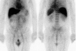
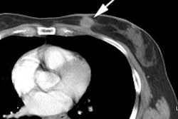
The agent was specific for myocardial necrosis, but is no longer commercially produced []. It binds irreversibly to exposed myosin filaments of damaged myosites. The antibody is targeted against the heavy chain of human cardiac myosin, a large intracellular protein in cardiac muscle cells that is exposed only when the integrity of the cell membrane is disrupted, resulting in irreversible injury. No adverse effects or allergic reaction have been reported. The agent also normally concentrates in the kidneys, liver, and less prominantly in the bone marrow. The critical organ is the kidney which receives an exposure of about 9 rads for a standard 2mCi dose. Focal non-antigen specific accumulation of the tracer has been reported at sites of lung inflammation and within soft tissue sarcomas. Diffuse non-specific pulmonary accumulation has been identified in post-cardiac transplant patients with PCP and CMV infections.
Antimyosin-Fab-DTPA-In111 is a Fab monoclonal antibody fragment, thus there is little or no risk of HAMA formation. No HAMA has been documented in any patient that has received one or multiple injections to date.
Antimyosin in Myocardial Infarction
The exam is positive as early as 1.5 to 4 hours post event, and can remain positive for up to 14 days. Images are best performed 24 to 48 hours after injection of the tracer (approximately 20% of activity remains in the blood pool at 24 hours). Antimyosin has an overall sensitivity of 92% for the detection of acute MI [1]. The sensitivity for transmural infarction is 94%, and 84% for non-transmural infarcts [2]. The extent of necrosis is an important prognostic indicator following myocardial infarction and the extent of antimyosin uptake correlates with the likelihood of experiencing re-infarction or sudden death. Activity at the site of an infarction fades over time, with about 50% of patients demonstrating decreased activity within 2 months. The duration of antimyosin uptake lasts about 6 months in most cases, and virtually all patients revert to normal in about 1 year. The abnormality associated with old MI's tends to be less intense than the abnormality associated with an acute MI because they contain only fibrous tissue.
Combined Myoscint/Thallium Imaging may provide prognostic information in the setting of the acute MI. A matched defect refers to an area of positive antimysin accumulation corresponding to a region with a fixed thallium defect. Such an area represents transmural necrosis/scar. These patients have completed their infarcts and there is no evidence of periinfarct ischemia. These patients are less likely to have a subsequent ischemic event.
An unmatched defect (ie: one in which there is both thallium and myoscint uptake) indicates that there is still viable myocardial tissue interspearsed with patchy or incomplete necrosis (or possibly subendocardial infarction) which may be at risk for subsequent ischemia. In fact, 20 of 28 patients with this pattern had subsequent ischemic events in one study [1].
Antimyosin in Unstable Angina
In one study, almost 50% of patients with unstable angina demonstrated focal uptake of the tracer. This suggests that many patients with unstable angina actually have areas of myocardial necrosis. Alternatively, this finding may represent the presence of a recent, but undetected small myocardial infarction in these patients. This finding has also been associated with recurrent ischemia with a sensitivity of 85% [2].
Antimyosin in Doxorubicin toxicity
In patients receiving doxorubicin, cardiac toxicity may produce irreversible and often fatal congestive heart failure. Continuous infusion of the agent has been associated with a reduced incidence of cardiotoxicity in comparison to bolus administration. In general, doses above 500 mg/m2 are associated with a greatly increased risk of cardiotoxicity, although it can be seen at doses less than this.
Intense antimyosin accumulation at intermediate cumulative doses (240-300 mg/m2) identifies patients at risk of cardiotoxicity before the ejection fraction deteriorates. Antimyosin is more sensitive to the detection of cardiac toxicity than a decreased EF and may help to identify patients who should receive alternate therapy.
Antimyosin in Cardiac Transplant Evaluation
Presently, frequent right heart biopsies are used to monitor heart transplant patients for rejection. Treatment is given for moderate or severe rejection, but withheld in patients showing only mild or no rejection.
Diffuse myoscint accumulation is seen in all heart transplants in the immediate post-operative period. This activity usually normalizes between 6 to 8 months in patients at low risk for rejection. In patients with serial studies, a declining heart to lung ratio (HLR) is associated with a benign clinical course (normal HLR is less than 1.5), but a persistently elevated or increasing HLR (HLR greater than 2:1) was associated with an increased risk for rejection complications. Care should be taken when determining HLR's as diffuse non-specific pulmonary accumulation of the tracer has been reported in post-transplant patients with PCP and CMV pulmonary infections. Increase pulmonary activity in these patients may falsely lower the calculated HLR. Persistent myoscint accumulation greater than 1 year post transplant is associated with a greatly increased risk for transplant rejection. It appears that antimyosin images may be a better predictor of future episodes of rejection than biopsy.
Antimyosin in Myocarditis
Histopathologic evidence of myocardial necrosis or lymphocytic infiltrates are found in only 10% of patients presenting with unexplained cardiomyopathy. Initiation of appropriate therapy for acute myocarditis idealy requires evidence of an acute inflammatory process. Biopsy is the definitive diagnostic tool, but this is not without risks, and sampling error make the test insensitive. In-111-Fab-DTPA imaging may be a useful screening tool to identify those patients requiring a biopsy. There is a fairly good correlation between positive antimyosin accumulation and a positive biopsy result. (Sensitivity 83-100% Specificity 50%.) Following treatment, about 50% of patients with positive antimyosin studies demonstrate improvement in their ejection fractions, whereas only 20% of patients with negative scans had improvement.
Antimyosin in Idiopathic Dilated Cardiomyopathy:
Uptake of antimyosin in commonly identified in these patients despite the fact the their biopsies are often negative for inflammation. This suggests that active myocyte damage is occuring in the absence of biopsy findings.
REFERENCES:
(1) Semin Nucl Med 1993; Manspeaker P, et al. Cardiovascular applications: Current status
of immunoscintigraphy in the detection of myocardial necrosis using antimyosin (R11D10)
and deep venous thrombosis using antifibrin (T2G1s). 23: 133-47
(2) American Journal of Cardiology, Cont. Ed. Series, Myocardial Perfusion Imaging, Part II (AJC, Part II)
(3) J Nucl Med 1995; Carrio I, et al. Indium-111-antimyosin and iodine-123-MIBG studies in early assessment of doxorubicin cardiotoxicity. 36: 2044-49
(4) J Nucl Cardiol 2009; Flotats A, Carrio I. Radionuclide noninvasive evaluation of heart failure beyind left ventricular function assessment. 16: 304-315


