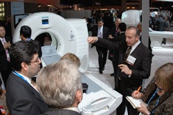
CHICAGO - Philips Healthcare of Andover, MA, is using its RSNA appearance to feature a new 128-slice CT scanner, as well as a 3-tesla MRI system. The company is also discussing a prototype PET/MRI system and a surgical C-arm based on flat-panel digital technology.
CT
In CT highlights, Philips is further diversifying its product portfolio with a 128-slice version of its iCT scanner, and the company is also bringing to North America a line of 16-slice systems currently sold internationally.
Brilliance iCT SP is a 128-detector-row system that gives customers a more value-oriented option to the company's flagship 256-slice iCT model. The scanner can also be upgraded to 256 slices as a facility's needs change, according to Philips.
The system sports a 0.27-sec gantry rotation speed and can be sited in 365 sq ft. It features Philips' Smart Focal Spot x-ray tube technology, which improves sampling density for enhanced spatial resolution in all exams, the company said.
Other features include the company's Nano-Panel detector design, which enables large area coverage for fast exams in clinical applications such as coronary artery imaging, lung scanning, and brain perfusion. Philips has also included its Eclipse DoseWise collimator on the scanner to reduce radiation dose.
The 16-slice product family, MX, is being displayed in North America for the first time, Philips said. The scanner is already sold internationally, and is ideal for customers who want to replace a current scanner or buy a second system but face economic constraints.
MX systems perform routine clinical applications such as CT angiography and are also appropriate for dental planning and virtual colonoscopy. The MX line is pending 510(k) approval from the U.S. Food and Drug Administration (FDA).
On the software side, Philips is highlighting new software applications for its Brilliance Workspace console, as well as Brilliance Everywhere thin-client software. The company is promoting automated features and new clinical applications for liver analysis, cardiac plaque assessment (shown as a work-in-progress), and whole-body bone removal for advanced vessel analysis.
The company is also touting its Perspective Filet View software for virtual colonoscopy studies. The application unfolds colon images, displaying the colon flat like a landscape in a 3D manner, which helps avoid "blind spots" created by folds. Philips claims the software can improve accuracy and reduce reading times for physicians.
MRI
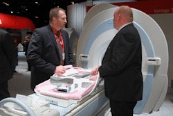
In MRI, Philips is launching a new version of its Achieva 3.0T TX, the newest generation of its 3-tesla MRI technology.
Achieva 3.0T TX includes a new multichannel radiofrequency (RF) transmission technology, called MultiTransmit, that adjusts the scanner's RF signal to patient anatomy. Philips believes that MultiTransmit is an improvement over single-channel transmit technology, addressing a number of the issues inherent in ultra-high-field scanning, such as dielectric problems and specific absorption rate (SAR). The end result is 40% faster image acquisition speed and image quality and more consistency in image results, according to the company.
 |
| Achieva 3.0T TX is the newest generation of Philips' 3-tesla MRI technology. |
At the 1.5-tesla field strength, Philips highlighting Achieva SE, a line extension of the company's Achieva 1.5T platform that's targeted toward more economy-minded customers.
Achieva SE uses the same basic platform as the high-end Achieve 1.5T A-Series scanner, and it includes the company's PowerSave technology to reduce energy consumption by up to 50%. The system also includes Philips' SmartExam protocols for improved workflow and image consistency. Achieva SE can also be upgraded.
In breast MRI, Philips is announcing the commercial availability of its Elite Clinical Solution for breast imaging, first introduced at the 2007 conference. The package includes techniques such as eThrive, MammoTrak dockable patient support, diagnostic and biopsy coils, and the DynaCAD software for analysis and biopsy planning from the MR console. The package is available as an option on Philips 1.5-tesla and 3-tesla scanners.
In other MRI news, Philips is releasing a 32-channel cardiac coil for Achieva 1.5-tesla and 3-tesla scanners, the extension of SmartExam to breast imaging with Breast Smart Exam, and more powerful gradients for the Panorama HFO high-field superconducting open scanner.
Molecular imaging
In oncology imaging, Gemini Big Bore PET/CT combines the Gemini TF PET system using TruFlight time-of-flight technologies with the Brilliance CT Big Bore system. A PET/CT can be performed in as few as five minutes. The system will be available in second quarter 2009.
The system incorporates a linear accelerator (linac) table and has an 85-cm bore diameter designed to facilitate more flexible patient positioning in radiation oncology cases. It also is better suited to accommodate large and obese patients. An Open View gantry space in the middle adds one-third more light, designed to help minimize patient claustrophobia.
Philips previewed the concept of an integrated PET/MRI scanner, the prototype models of which are being installed at research sites. This in-development system fuses MRI and PET images for use in molecular imaging applications.
The new BrightView XCT, shipping in second quarter 2009, combines the BrightView SPECT in a coplanar design with advanced Philips flat-detector x-ray CT technology. The system uses a flat-panel x-ray detector for the purpose of anatomic localization and attenuation correction in nuclear medicine. Coplanar SPECT and CT capabilities eliminate the need to index the bed between the two acquisitions.
Mammography
Philips has been eyeing the full-field digital mammography (FFDM) market segment in recent years, and this year the company is once again showing RSNA attendees its MammoDiagnost DR system. The unit is not yet available in North America, and Philips is preparing a regulatory submission to the FDA.
Also on display is MammoDiagnost Vu, a standalone mammography workstation that enables users to view images from FFDM systems from multiple vendors. The workstation can also automatically align the left and right breast tissue according to the specified hanging protocol. MammoDiagnost Vu is pending 510(k) clearance and is being shown as a work-in-progress.
Philips is also discussing mammography applications on its PCR Eleva CosimaX computed radiography (CR) system, which can be used with existing analog mammography units, giving a mammography facility an economical entrée into digital mammography. Mammography is not yet available on PCR Eleva CosimaX in the U.S.
Finally, Philips is discussing the integration of SecondLook Digital software from iCAD of Nashua, NH, on its digital radiography (DR) and CR mammography systems. The software is not yet available on Philips systems in North America.
Healthcare informatics
Philips is demonstrating iSite Radiology and iSite Enterprise PACS with the latest versions of its advanced visualization tools. These tools empower any physician who has access to an iSite network with advanced visualization functionality. Philips anticipates that by making advanced visualization capabilities available to all users, workflow delays, generated, for example, by the need to obtain a replacement or additional 3D projection, will be minimized.
For power users requiring the most sophisticated 3D advanced visualization functionality, an updated iSite Volume Vision software package may be added to diagnostic workstations. Its seamless integration with Philips enterprise architecture eliminates the need to purchase a third party advanced visualization system, and this may be its greatest appeal. The seamless integration also eliminates the costs associated with routine operation, maintenance, and space that would be required for a separate server.
Philips is underwriting a "closed loop" imaging research trial with the University of Chicago Medical Center. Its objective is to find the most effective ways to integrate information systems with imaging systems by identifying and removing bottlenecks and inefficiency at all points within the imaging and reporting process. The findings and solutions of this trial are expected to be productized into iSite. Not surprisingly, the project is under the direction of Dr. Paul Chang, medical director of enterprise imaging at the University of Chicago Hospitals. Chang was the founder of Stentor, the company that commercially developed the iSite PACS.
Pulmonary embolism assessment software and high-end CT colonoscopy software for use with iSite, both works-in-progress, are making their preliminary debut at RSNA 2008. The 3D colonoscopy visualization displays four tiles of images that represent an "unfolded cube" of an image in a colonoscopy fly-through. This visualization format is designed to enable greater scrutiny of details on an image-by-image basis.
X-ray
The next generation of Philips' Allura Xper line of interventional imaging systems is taking center stage in the company's booth. The new Allura family is designed to support applications from neuro to abdominal to peripheral and comes in monoplane and biplane configurations, along with a set of 3D interventional tools.
The company is highlighting a new Release 7 version of the Allura Xper FD20 system, which includes a remote servicing option that enables Philips engineers at the company's service headquarters to review exam protocols remotely, as they are being conducted. Philips is also showing Xper FD20 with a 56-inch 8-megapixel monitor designed to provide better review of images during procedures.
The company is addressing concerns about radiation dose to hospital staff by demonstrating Dose Aware, a new concept in dose monitoring that enables users to view dosimetry information in real-time. Personnel in the angiography suite wear special dosimetry badges that transmit data to a monitor within the suite; the monitor displays dose rising or falling depending on the location of staff, enabling them to take action immediately to lower their radiation exposure. Dose Aware is being shown as a work-in-progress and will be available as part of the Release 7 package.
Philips is also touting XperGuide, which provides live 3D needle image guidance, bringing percutaneous needle interventions back to the angiography lab. The tool creates an overlay fluoroscopy and XperCT 3D soft-tissue imaging that provides information on needle path and target. The company said that XperGuide can be used for applications from biopsies and drainages to RF ablations. New features include the ability to accept CT or MRI data for planning.
Another application, Dynamic 3D Roadmap, provides live 3D guidance through tortuous vasculature by creating an overlay of real-time 2D fluoroscopy images and the 3D reconstruction of the vessel tree. Philips believes the application can help improve visualization during complex interventions, including neuro aneurysm and uterine fibroid embolization.
Philips is also featuring its Allura 3D-RA rotational angiography application for 3D visualization of the brain, peripheral vessels, and bone structure, and XperCT, which brings CT-like images to the angio suite, with reconstruction times as low as 25 seconds and acquisition frame rates up to 60 frames per second.
Finally, Philips is highlighting Xper Table, which offers automatic position control to increase accuracy and improve workflow in interventional procedures, and Xper Cradle, which allows interventionalists and surgeons to roll patients while keeping the region of interest in isocenter.
On the software side, Xper Information Management Vascular Monitoring 5 is a new electronic charting, procedural monitoring, and report generation application for interventional and invasive radiology suites.
In radiography and fluoroscopy, Philips is promoting its Eleva Wireless flat-panel digital radiography detector. The detector has been designed for Philips digital x-ray systems and will initially be integrated into the company's DigitalDiagnost radiography rooms. The detector is pending 510(k) clearance.
The newest release of DigitalDiagnost includes an optimized workflow thanks to the use of the Eleva user interface on the system. The unit also includes Philips' Unique image processing software.
Other Philips x-ray highlights include the economy-priced Essenta DR system with a U-arm geometry, the EasyUpgrade DR program, the PCR Eleva computed radiography line, and xLNA Enterprise computer-aided detection software for evaluating pulmonary nodules.
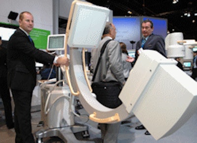
In interventional imaging, Philips is touting 3D imaging on its MultiDiagnost Eleva 3D-RX system. For the company's BV Endura and BV Pulsera surgical C-arms, Philips is offering a look at its SmartVision technology for producing high-quality images at low radiation dose.
 |
| Veradius is a new flat-panel digital surgical C-arm. |
Ultrasound
In the world of ultrasound, a new upgrade, Vision 2009, incorporates tissue aberration correction technology and algorithms. This is part of an initiative to better define tissue in fatty breasts. Tissue aberration correction, which was devised initially to look at obese patients, compensates for speed of sound variations in dense tissue, improving detail resolution and conspicuity of lesion details.
Ambient Experience
Introduced in 2004, Philips is expanding its imaging suite room environments, which are designed to provide simple, efficient work environments for imaging professionals and a soothing ambience for patients. This year, Philips demonstrated a PET/CT imaging suite, incorporating a patient prep room, PET/CT suite, and control room. Patients select the room theme of their choice using a mobile tablet in both the prep room and the procedure room. Themes include children-oriented designs.
The Ambient Experience concept has also expanded to emergency departments, the first of which has been, perhaps appropriately, installed at the newly constructed Disney Children's Hospital on the campus of Florida Hospital in Orlando.
Healthcare finance
The global economic healthcare crisis has been crimping the ability of a number of hospitals and imaging centers to obtain financing for capital equipment purchases, and Philips is promoting its Philips Medical Capital (PMC) arm as an option. PMC is a joint venture between Philips Healthcare and financing firm De Lage Landen, a division of Dutch banking giant Rabobank. PMC can provide customers with fixed-rate financing at predictable rates, the company said.
By Brian Casey and Cynthia Keen
AuntMinnie.com staff writers
December 3, 2008
Related Reading
Philips names new CMO, November 30, 2008
Philips Healthcare cuts jobs by 5%, November 21, 2008
Philips teams with NeuroNexus, November 17, 2008
Road to RSNA, Healthcare Informatics, Philips Healthcare, November 10, 2008
Road to RSNA, Archiving Systems, Philips Healthcare, November 7, 2008
Copyright © 2008 AuntMinnie.com