CT angiography (CTA) using moderate doses of IV contrast in patients with advanced renal failure is a safe procedure that negatively affects renal function in only a small percentage of patients, according to researchers from Baltimore.
But the imaging options are even better with newer MDCT scanners. CTA protocols with low kVp and using half the normal dose of iodinated contrast have even less impact on kidney patients and produce excellent images, said Dr. Barry Daly from the University of Maryland.
In a study presented at last month's International Society for Computed Tomography (ISCT) meeting in San Francisco, Daly showed how even normal contrast doses had little effect on serum creatinine levels in most patients. Information gained by the studies far outweighs the chance of adverse effects in patients with chronic renal failure, he said.
Daly also showed his latest protocol for low-contrast-dose, low-kV CTA imaging that delivers high image quality with even less risk for these patients.
Don't skip the CTA
CTA isn't something you want to skip, even though many centers do just that, he said. Before renal transplant surgery, for example, surgeons need to see what they're going to be dealing with in the operating room.
"There are big risks going into surgery without CTA in this group because they have the risk of a major change in operative procedure, prolonged surgery, poor graft outcomes, failure to engraft, loss of the organ -- which is a total disaster, especially in the setting of matched renal donor transplantation -- and, of course, the possibility of missing important pathologies," Daly said.
 |
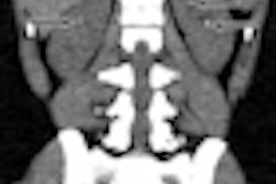
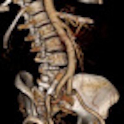
| Patient is in renal failure but not yet on dialysis. CTA was acquired at 80 kVp and 360 mAs following administration of 50 mL of 350 mg/mL contrast. Contrast density in the iliac arteries is > 400 HU. Radiation dose is 4.3 mSv. There is an incidental right iliac venous stent. Images courtesy of Dr. Barry Daly. |
 |
Daly and colleagues reviewed the use of CTA in 180 potential renal transplant recipients, all with matched donors, "but we were especially interested in looking at a predialysis cohort of 40 patients," he said.
Patients were assessed for aortoiliac and calcific atherosclerosis, venous thrombosis, and increased incidence of renal cell carcinoma (four to seven times the normal rate in native kidneys). The only prophylaxis was aggressive oral hydration both before and after CTA, he said.
It's important to find an appropriate place for the engraftment, Daly said. Many patients may require surgical correction with bypass grafts before transplantation can be done.
"Because a lot of these folks have had chronic hemodialysis, it's not uncommon to find occlusion of the iliac veins," he said. In a couple of cases, this has become obvious only in the operating room -- "with disastrous consequences."
The study measured serum creatinine (SeCr) and estimated glomerular filtration rate (eGFR) before and after imaging in the 40 predialysis patients. "There was almost no difference between the two groups in mean measurements," Daly said.
"Nobody had to undergo dialysis, but looking at the changes, there clearly were some shifts that we shouldn't ignore," he said.
Post CTA, 27 patients had stable or mean decreased SeCr of 10.2%. Thirteen patients had a mean increase of 6.3%, with one patient showing a 32% rise, and three saw almost no change (0.5-1.5 mg/dL increase).
Change in SeCr after CTA in predialysis cohort of 40 patients
|
A low rate of contrast-induced nephropathy (CIN) in renal patients isn't all that surprising, Daly said. Newhouse, Katzberg, and others have shown that the risks of IV low-osmolar contrast precipitating CIN have been overestimated.
These previous studies demonstrate that moderate doses of IV contrast are much less nephrotoxic than arterial administration, he said. Many studies without controls failed to allow for other factors affecting renal function, especially in hospital populations. Finally, a 2010 study in Radiology showed that low-osmolar contrast may be as safe as iso-osmolar contrast, he noted.
Low-kVp, low-contrast-dose CTA
Daly and colleagues also performed a study of low-kVp, low-contrast-dose CTA in chronic renal failure patients. Why is this necessary if the regular dose is safe?
"The answer, of course, is that lower is always better," Daly said. Even if the negative effects aren't as bad as they were feared to be, "there is still a small portion of patients in our group who are still at risk with the conventional dose," he said, adding that "new CT scanners have enabled new techniques for getting more out of each gram of iodine."
The technique involves dropping the kV and increasing the mAs. For example, if you drop the tube current from 120 kV to 80 kV, you would increase the mAs by a factor of 2.7. Thus, 120 kVp and 250 mAs become 80 kV and 600 mAs, he said, and for large patients the tube may reach the maximum mAs.
MDCT can be used with extended z-axis coverage to shorten scan times to correspond to a shorter bolus train. Ideally, there should be at least 40 mm to 80 mm of coverage, he advised.
How it works is by now well-known, he said. "The k-edge of iodine is only [33.2] keV, so by dropping our kVp we can actually get considerably increased x-ray absorption. There's a nearly a twofold increase in iodine attenuation at 80 kV compared to 120 kV." Thus, the iodine dose can be cut in half while producing similar CT values, according to Daly.
The researchers perform the 80-kV studies on a Brilliance iCT 256 or a Brilliance 64 scanner (Philips Healthcare) using 320 to 350 mgI/mL of contrast.
They inject 35 to 50 mL of contrast at 4 mL per second in a peripheral vein, followed by 40 to 60 mL of a saline chaser at 4 mL per second. Automated bolus tracking is set for a 120- to 150-HU threshold in the aorta just below the hiatus, and the automated minimum scan delay is set to 4.2 to 6.5 seconds, he said.
Tube rotation speed is 0.75 seconds for a 256-slice scanner and 0.75 to 1 second for a 64-detector-row scanner. mAs values are based on patient body mass index (noise present at precontrast phase) with pitch set at maximum for the mAs selected, Daly said.
The resulting images look great, but using iterative reconstruction (iDose5, Philips) allows even lower doses, or the scanning of larger patients using low-kV, low-contrast protocols, he said.
In summary, CTA with a moderate 100-mL dose of iso-osmolar contrast in advanced renal failure "is a safe procedure with a negative impact on renal function in only a small percentage of patients," Daly said, adding that "these aren't patients with creatinine of 1.8, these are patients with major renal compromise."
However, even though the 100-mL contrast protocol has a "very limited negative effect on people ... a better option today is low 80-kVp, low-contrast-dose CTA technique with 35 to 50 mL of iso-osmolar contrast," Daly said.
This very safe technique yields high diagnostic quality and works on most newer CT scanners, he added.
"Finally, if you have the benefit of iterative reconstruction, it improves image quality and allows us to use this technique even in very large patients," Daly said.