Pediatric radiation dose was cut by as much as half for some clinical applications by swapping 64-detector-row CT for 320-detector-row scanning, according to researchers from Cincinnati Children's Hospital Medical Center. The study indicates that advances in CT technology can be successfully used to cut pediatric radiation dose.
The study, on phantoms, showed significant dose reductions that varied significantly depending on the scan region with a volumetric 320-detector-row CT scanner. There were other potential benefits of wide-area scanning as well, the group found. The research was presented at the Society for Pediatric Radiology (SPR) meeting in San Francisco earlier this month.
"There are potential dose savings associated with volumetric scanning compared to helical scanning: For instance, because there's no need to extend the scan length beyond the image boundaries in order to reconstruct the first and last slices, excess dose from overranging is eliminated," said Dr. Jennifer Johnston. "There are also no overlapping helical sections and there's less overbeaming due to the smaller penumbra."
Many studies have shown outsized growth in CT utilization in recent years, including in pediatric hospitals; a recent study showed a 4.8-fold increase from 1995 to 2008, Johnston said. Children, of course, face unique physiologic risks from exposure to ionizing radiation due to their longer life expectancy and higher cell division rate compared to adults. One potential path to lowering radiation exposure is through advances in CT technology.
Scanning with 320-detector-row CT provides 16-cm z-axis coverage in a single 0.35-sec rotation. Other available scan modes on the system include conventional helical scanning with 64 detector rows, fast ultrahelical scanning with 160 detector rows and 8-cm coverage per rotation, and volume scanning with 320 detector rows. The increased z-axis coverage in wide-area scanning enables faster scans with less motion artifact and less need for sedation in children, Johnston said.
"There is a lack of accurate pediatric volume dosimetry data in the literature, and there's no literature to our knowledge comparing 320-detector-row helical with 64-detector-row CT for pediatric abdominal imaging," she said.
The study by Johnston, Dr. Daniel Podberesky, and colleagues compared the three scan modes for body imaging on a 320-detector-row scanner (Aquilion One, Toshiba America Medical Systems) with regard to organ doses, effective dose, acquisition time, and image noise. They compared the three scan modes in three abdominal regions: chest, chest/abdomen, and chest/abdomen/pelvis.
Conventional 64-detector-row helical scans were acquired both on the 320-detector-row machine and on a 64-detector-row scanner (Aquilion 64, Toshiba). The scanning parameters were designed to be similar to each other, with the same tube voltage, automatic exposure control, reconstruction kernel, and noise reduction algorithm, and all settings represent currently used clinical protocols, Johnston said.
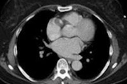
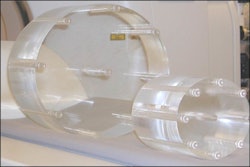
Radiation exposure was measured in an anthropomorphic phantom representing a 5-year-old child made of tissue-equivalent materials and equipped with metal-oxide-semiconductor field-effect transistor (MOSFET) detectors calibrated to the scanner type. Noise was measured at four levels for each protocol; effective dose and noise were compared with a paired t-test or sign test.
Effective dose was calculated for each protocol using International Commission on Radiological Protection (ICRP 103) tissue-weighting factors and then summing the weighted organ doses.
All doses lower at 320
"The effective doses contained on all 320-detector-row CT modes were all statistically lower than those obtained on the 64-detector CT," Johnston said.
The highest organ doses were in the chest (adrenal glands/kidneys) at 5 mGy at 64-detector-row CT. In the abdomen/pelvis and chest/abdomen/pelvis, the highest organ doses were in the small (10 mGy) and large (11 mGy) bowel, again at 64-detector-row CT.
 |
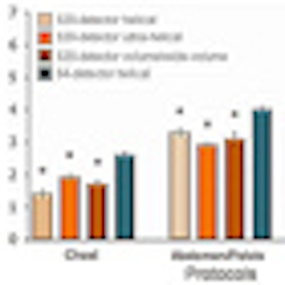
| Effective doses for each scan mode in the study, divided by the body segment scanned. Effective doses for scans performed on 64-detector-row CT were significantly higher than those for all scan modes on 320-detector-row CT. Data and images courtesy of Dr. Jennifer Johnston. |
The maximum dose savings that could be achieved with 320-detector-row CT were as follows:
- Chest: 64 detector (2.6) versus 320 helical (1.4) -- dose savings 46%
- Abdomen/pelvis: 64 detector (4.0) versus 320 ultrahelical (2.9) -- dose savings 28%
- Chest/abdomen/pelvis: 64 detector (5.9) versus 320 helical (5.1) -- dose savings 14%
There were no significant differences between effective doses among the three scan modes evaluated on 320-detector-row CT.
Image acquisition times at 320-detector-row CT were shorter than all results at 64-detector-row CT, she said. The differences between helical modes were not great; larger differences were seen between helical and volume or wide-volume modes. But the greatest observed difference was seen in chest imaging, comparing volume mode with helical mode on a 64-detector-row CT.
 |
| Scan durations for all protocols performed on 320-detector-row CT were shorter than those performed on 64-detector-row CT. Although the difference between helical modes was small, there were greater differences when comparing ultrahelical and volume or wide-volume mode to helical mode on the 64-detector-row CT, with the greatest observed difference in chest imaging between volume mode and helical mode. |
Image noise was similar across scans overall, although there were some statistically significant differences with volume mode on chest imaging and wide-volume and ultrahelical modes for abdomen/pelvis and chest/abdomen/pelvis.
"However, the differences overall were not greater than a few step deviations of Hounsfield units, and it's uncertain what the clinical significance of this finding is," Johnston said.
Among the study's limitations, only a single phantom size was tested, and effective dose is an imperfect descriptor of dose. Finally, there is more to image quality than just image noise, she said.
Significant dose savings can be achieved in pediatric body CT on a 320-detector-row CT compared to a 64-detector-row scanner, Johnston concluded. Shorter scan durations with 320-detector-row CT also potentially lead to fewer motion artifacts and less need for sedation.