When using CT to assess graft patency after coronary artery bypass graft (CABG) surgery, employing a variable-pitch technique speeds scanning and reduces contrast dose by a third, Japanese researchers said in a presentation earlier this month at the RSNA meeting in Chicago.
"Bypass grafts extend all the way from the subclavian artery to the coronary arteries, significantly increasing imaging time," explained Toshihiro Ishihara, MD, from Chiba Medical Center in Chiba, Japan. "As a result, complicated imaging procedures are employed, such as scanning regions of the bypass graft separately, or scanning a large area with [electrocardiogram (ECG)] gating. However, using variable-pitch CT, it's possible to switch from non-ECG-gated scanning to ECG-gated scanning in a single continuous imaging procedure."
A proprietary variable helical pitch CT scanning technique (vHP, Toshiba Medical Systems, Otawara, Japan) works by varying the table speed while scanning to obtain continuous images during a short scan in a single phase. The heart is scanned using a narrow pitch with ECG gating, and the abdomen is scanned using a wider pitch and no gating, Ishihara said.
The study aimed to assess the coronary arteries and post-CABG patency "in order to determine whether the volume of contrast can be reduced while maintaining acceptable CT numbers," Ishihara said in his presentation.
In all, 31 patients underwent CT assessment of graft patency following CABG:
- Twelve patients (10 men, two women; mean age, 66.8 years; range, 53-77) were scanned without variable helical pitch (non-vHP group).
- Nineteen patients (13 men, 6 women; mean age, 67 years; range, 63-75) were scanned with variable helical pitch (vHP group).
Nonhelical dynamic scans were acquired at 120 kV and 300 mA (0.35-sec rotation) using a 64-detector-row CT scanner (Aquilion 64, Toshiba). Helical scans were acquired at 120 kV and 600 mA (0.35- to 0.45-sec rotation).
Contrast agents iopamidol 370 or iopremol 350 were injected at a rate based on body weight multiplied by 0.7 mL/sec and followed by a saline solution flush, and ECG-gated scanning was performed to generate axial images using the retrospective gating method, the authors noted in an accompanying abstract.
- For the actual scans following a test scan, the contrast medium volume was equal to the injection rate (mL) multiplied by 10 (scan time).
- The saline flush (mL) was equal to the body weight multiplied by 0.25.
- The scan start time (seconds) was equal to the peak enhancement time plus two seconds.
The team then measured total contrast volume and the HU value in the aorta at the origin of the left main trunk (LMT) to evaluate the effects of using variable helical pitch.
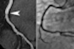
"Graft patency could be accurately assessed in all patients," Ishihara said.
The patient group without variable helical pitch used an average contrast medium injection volume of 71.7 mL, and the average HU value in the aorta at the origin of the LMT was 526.9 HU.
Comparison of non-vHP and vHP groups
|
With vHP scanning, the volume of contrast media injected could be reduced by approximately 33%, while maintaining CT values not significantly different from those in non-vHP scanning, Ishihara said.
The contrast protocol is extremely important for the shorter vHP scans, he said, as it must begin and end while the contrast is in the vessels. Patient factors such as heart rate and characteristics of the system must therefore be incorporated into the protocol.
The use of vHP scanning can significantly shorten the scan time and, hence, contrast volume, which is "of great value in the clinical setting, with many benefits such as reducing the risk of contrast-induced nephropathy," Ishihara said.
By Eric Barnes
AuntMinnie.com staff writer
December 20, 2010
Related Reading
Automated PE protocol tool optimizes contrast use, image quality, November 29, 2010
CCTA image quality drops at low doses in big patients, October 12, 2010
Few adverse events occur a year after negative cardiac CTA, September 21, 2010
Body mass index predicts left atrial thrombus in atrial fibrillation, December 31, 2009
Timing is everything in contrast-enhanced CT, September 28, 2006
Copyright © 2010 AuntMinnie.com