Researchers at the University of Vermont in Burlington showed promising results of how parallel transmission (multitransmit) MRI at 3-tesla magnet strength can improve image quality in the lumbar spine by reducing shading and other dielectric effects.
The study, presented last week at the annual 2010 RSNA meeting in Chicago, also reduced scan time for patients using the multitransmit technique.
"We have found, at least anecdotally, that when we image people in their spines, there are often issues that we have with dielectric shading on the edges of the field," said Christopher Filippi, MD, director of MRI, section head of neuroradiology, and medical director of the university's Biomedical Imaging Center. "In particular, it is seen often in patients with large body habitus and equally in patients who are very fit and with a single percentage of body fat."
Improving S/N and C/N ratios
Multitransmit MRI uses radiofrequency (RF) coils in which the frequency, amplitude, phase, and waveform of all RF sources are automatically adjusted for optimal uniformity for each patient's anatomy. Filippi and colleagues wanted to determine if the multitransmit MRI technique would improve signal-to-noise (S/N) and contrast-to-noise (C/N) ratios in lumbar spine MR imaging at 3 tesla.
The study enrolled a total of 19 volunteers. Nine men and one woman with an average age of 30.7 years, ranging from 16 to 47 years old, received T1-weighted MRI lumbar scans. In addition, nine healthy volunteers (six men and three women) with an average age of 30.2 years, ranging from 16 to 46 years old, received T2-weighted MRI lumbar scans.
The researchers also performed background noise scans with no volunteers in the magnet to calculate S/N and C/N ratios. They then proceeded with routine conventional single-transmit MR and multitransmit MR scans on the volunteers. Sagittal and axial T1- and T2-weighted images were acquired using both multitransmit MR and conventional MRI.
The study also divided the volunteers into two groups for the sagittal and axial MR imaging, because it would take more than two hours of scan time to image a patient. "No normal volunteer is going to tolerate over four hours of imaging research in a magnet," Filippi added.
Image evaluation
Percent improvements in S/N and C/N ratios were calculated, and reductions in scan time also were noted. With the help of three neuroradiologists, the researchers evaluated the percentage of improvement in the scans, based on a simple three-point scale of better, worse, or the same.
"This is what we did for every slice in the sagittal and axial planes on all 19 people," Filippi said. "It takes hundreds of hours to calculate this, but we were really obsessional."
The analysis found that for sagittal T1-weighted MR images, C/N with multitransmit MRI improved by 53% and S/N by 19%. For axial T1-weighted images, C/N rated a 48% improvement, while S/N was enhanced by 23%.
 |
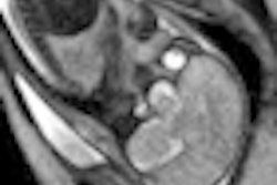
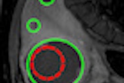
| Images of the lumbar spine show reduced shading and improved C/N between a standard MRI (left) and multitransmit MRI (right) with T1-weighted imaging. All images courtesy of Christopher Filippi, MD. |
For sagittal T2-weighted images with multitransmit MRI, C/N improvement was 38% and S/N was 20%. For axial T2-weighted scans with multitransmit MRI, C/N improved by 18%, while S/N improved 100%.
 |
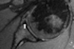
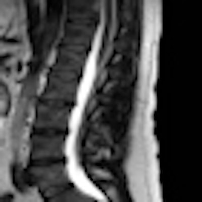
| The lumbar spine shows reduced shading and improved image quality when comparing standard MRI (left) and multitransmit MRI (right) with T2-weighted imaging. |
The researchers also found a reduction in scan time of 35% on sagittal T1- and T2-weighted imaging, while a 50% reduction in scan time was achieved on axial T1- and T2-weighted imaging.
Most important, multitransmit lumbar spine MR image quality was judged "superior" to routine MRI in all cases by two neuroradiologists and equal to or better in all cases by the third neuroradiologist.
"It was important that we proved that these images were better. The next step is: Can we actually have better lesion detection? I don't know if the answer is out there yet," Filippi said. "Certainly, when we work with people who have obesity and people who are really thin, it is hard to image them well. So, maybe this will be a great help to us, but we need to do that study next."
The researchers also plan to determine if there are similar imaging improvements in the cervical and thoracic regions using the multitransmit MRI technique.
By Wayne Forrest
AuntMinnie.com staff writer
December 9, 2010
Related Reading
Stand-up MRI detects impact of heavy backpacks on children, January 6, 2010
MRI reveals high incidence of disk disease in overweight and obese youngsters, December 2, 2009
MRI finds proximal femur fractures missed by x-ray, September 1, 2009
MRI helps find fractures among elderly female ED patients, April 29, 2009
Copyright © 2010 AuntMinnie.com