CHICAGO - CT imaging, the most commonly used neuroimaging procedure to monitor children with shunted hydrocephalus, also puts these patients at high risk of developing a radiation-related fatal cancer as adults. MRI exams using rapid brain protocols can produce diagnostically acceptable results and eliminate the risk from repeated exposure to CT radiation dose.
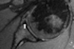
In addition to not subjecting children -- especially very young children -- to radiation dose exposure, rapid MRI of the brain has been proved satisfactory for assessing ventricular size, and it does not require sedation because image acquisition can be completed in 20 seconds.
Pediatric radiologists from Children's Medical Center in Dallas made this MRI recommendation to RSNA attendees earlier this week, while delivering a sobering assessment of the cancer risk they believe their patients would face if head CT exams were continued until the children reached 20 years of age.
In a discussion of the risks of CT, it was acknowledged that the lifetime cancer risk of having a head CT scan is considerably less than with other types of CT exams, especially when low-dose protocols are utilized. Korgun Koral, MD, an associate professor of pediatric radiology at the University of Texas Southwestern Medical Center in Dallas, addressed the subject.
A 3-year-old girl has an eight in 10,000 mean lifetime risk of developing a radiation-related cancer from a head CT exam, compared to 40 in 10,000 odds with a chest CT exam. Also, the older the patient, the less the radiation dose risk. The risk of radiation-related cancer from a head CT exam is halved for a 15-year-old girl, and is halved again at age 30. (For boys, the risk is nine in 10,000, five in 10,000, and three in 10,000 at ages 3, 15, and 30, respectively.)
However, comparative assessments do not diminish the risks to a pediatric patient with shunted hydrocephalus. These patients typically have head CT exams several times a year, usually beginning at very young ages. Hydrocephalus, the abnormal accumulation of cerebrospinal fluid in the brain, affects one in every 500 children, according to the National Institute of Neurological Disorders and Stroke (NINDS) in Bethesda, MD.
To treat the condition, which may be congenital or acquired, shunt systems are used to drain the cerebrospinal fluid. Treatment improvements have increased the life expectancy of these patients, Koral said.
Koral and colleagues conducted a retrospective study to estimate the lifetime attributable risk of children with shunted hydrocephalus developing a fatal cancer due to head CT for ventricular size assessment. They reviewed the medical records of Children's Medical Center from January 2009 through March 2010.
The researchers identified 150 patients, 81 of whom were female. The average age of the patients was 1.76 years, although ages ranged from 1 day to almost 19 years. To calculate the average number of exams per year that each patient underwent, the researchers counted neuroimaging studies residing in the hospital's PACS archive that were performed on these patients. In total, 211 rapid brain MRI exams and 910 head CT exams were identified. They found that 85% percent of the CT exams were performed between 6:00 a.m. and 9:00 p.m., when MRI technologists were on duty at the hospital and MRI exams could have been scheduled instead.
On average, four neuroimaging studies were performed each year, and the researchers made the assumption that these studies began for a typical patient in the second year of life. The lifetime attributable risk of developing a fatal cancer was calculated with the assumption that a patient would have four exams per year through the age of 20.
"At our hospital, the low-dose CT protocol used for hydrocephalus evaluation is 1.1 mSv, and if a standard CT protocol is used, 2.5 mSv," Koral said. "If a low dose is used consistently throughout an 18-year period, the risk of developing a fatal cancer is one in 124. If the standard dose is utilized, the risk is one in 52 patients."
"What this means is that in our current patient population, between one and three children will develop a fatal cancer if they survive to live an average adult lifetime," he said.
The researchers determined these risk estimates based on the Biologic Effects of Ionizing Radiation (BEIR) VII report, and effective doses obtained using the International Commission on Radiological Protection (ICRP) Report 103 organ weighting factors.
Whenever possible, the children's hospital uses rapid brain MRI to assess ventricular size of its patients with shunted hydrocephalus.
By Cynthia E. Keen
AuntMinnie.com staff writer
December 3, 2010
Related Reading
Pediatric CT settings should be adjusted to child's body size, May 25, 2010
Color-coded CT protocols help reduce pediatric radiation dose, June 4, 2009
SPR news: Rads must take lead in reducing pediatric CT dose, April 23, 2009
ARRS study: Child's body shape can reduce CT dose, April 23, 2009
Copyright © 2010 AuntMinnie.com