CHICAGO - FDG-PET/CT is useful in the early diagnosis, follow-up, and management of post-transplant lymphoproliferative disease (PTLD), according to a University of Nebraska study presented Wednesday at the 2010 RSNA conference.
The study, led and presented by Homeira Zahiri, MD, from the University of Nebraska Medical Center in Omaha, also concluded that the clinical relevance of using FDG-PET/CT in the management of PTLD should be evaluated in a larger prospective cohort to confirm its efficacy.
PTLD is a severe complication that may occur any time after solid organ or bone marrow transplantation. According to previous research, it is the second most common malignancy in adult recipients and the most common post-transplant malignancy in children. Other research has found that the five-year cumulative incidence of PTLD for pediatric recipients is 3.2%, compared with 0.9% in adults.
Modality comparison
While ultrasound, MRI, and CT are the most commonly used imaging modalities in PTLD, Zahiri noted that there are no large studies comparing the various modalities in PTLD applications.
Ultrasound is useful in identifying changes in solid organs, such as in liver and kidney transplants, and can detect the changes due to PTLD within the abdomen in some cases, Zahiri said. MRI is helpful in diagnosing bone involvement and central nervous system lymphoma.
"CT is helpful in assessing the extent of disease, identifying area for biopsy, staging, and treatment response," she added. "It is incapable of identifying the involved nodes that have not increased in size and is very poor at identifying bone involvement, which would automatically put a patient into stage IV of the disease."
 |
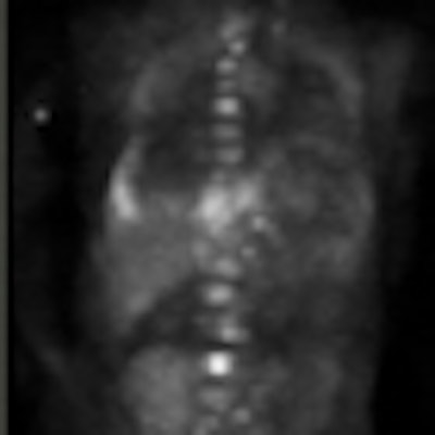
| The patient in this PET image had repeated CT scans with inconclusive findings. FDG-PET discovered extensive abnormal bone and liver radiotracer uptake, which was confirmed as PTLD by biopsy. Image courtesy of Homeira Zahiri, MD. |
FDG-PET/CT benefits
Based on prior research, Zahiri said the prime role for FDG-PET/CT would appear to be in assessing response to therapy, rather than aiding in diagnoses.
In this study, Zahiri and colleagues retrospectively analyzed 12 patients from the University of Nebraska Medical Center with histologically confirmed PTLD and who had positive FDG-PET/CT exams. Seven patients had at least one follow-up FDG-PET/CT scan after treatment.
In addition, the researchers retrospectively reviewed all CT, MRI, and PET images of all 12 patients and correlated the findings on a lesion-by-lesion basis. The analysis of the imaging data included the patient's initial staging of PTLD and the response to therapy.
In analyzing the data, the researchers found that at the initial staging of PTLD, all sites of disease involvement as detected by conventional imaging also were identified through areas of abnormal radiotracer uptake by FDG-PET/CT.
Staging PTLD
In addition, the researchers found no difference between anatomical and functional imaging at staging of disease in six of the 12 patients. "However, in the other six patients, CT -- and in one case, MRI -- was not able to detect bone involvement, whereas FDG-PET showed significantly extensive pathological tracer uptake, which upstaged [PTLD] from stage II and stage III to stage IV of the disease," Zahiri said.
In conclusion, Zahiri noted that the findings agreed with previous observations that FDG-PET is "superior" to conventional imaging methods for detecting extranodal localization.
"FDG-PET performed after treatment may provide a more accurate response classification and prediction of prognosis as compared to CT-based assessment by distinguishing viable tumor from necrosis and fibrosis in residual mass," Zahiri said. "Therefore, FDG-PET is useful for staging and management of PTLD, with a great potential for response after therapy."
By Wayne Forrest
AuntMinnie.com staff writer
December 1, 2010
Related Reading
Test may let transplant patients skip biopsies, April 22, 1010
CT maintains edge over MRI in liver transplant planning, March 10, 2008
CT measures post-transplant bronchiolitis obliterans severity, March 14, 2006
FDG-PET may predict which lymphoma patients will benefit from stem cell therapy, December 30, 2005
Copyright © 2010 AuntMinnie.com