CHICAGO - Radiologists should describe superior and posterior labral lesions as fraying and/or tearing in their reports to surgeons, unless radiologists can clearly see a displaced tear, according to a study presented Monday at the RSNA annual meeting.
The information gleaned from MRI results can provide preoperative information for the surgeon. The radiologist can alert the surgeon to a labral tear that is displaced and communicate whether it's a surgical lesion or a nondisplaced tear that may not require surgery.
The purpose of the study "came about as part of our quality assurance program in our practice in discussions with orthopedic surgeons and their perception on how often we are calling certain labral lesions versus other lesions," said lead study author Thomas Magee, MD, managing partner at Neuroskeletal Imaging in Merritt Island, FL.
The retrospective study included 100 consecutive patients who had shoulder labral tears described on MR examination and who went to subsequent surgery. Surgical reports were correlated with MR examinations.
Patient population
The average age of the patients was 39 years, ranging from 14 to 72. Most of the MRI scans were performed on a 3-tesla system (GE Healthcare, Chalfont St. Giles, U.K.), with a standard imaging protocol performed on all 100 patients. Some patients received both an MRI scan and an MR arthrogram.
"It was specifically determined if surgical intervention, such as surgical tracking of debridement, was performed on the lesions described by MR exam," Magee added.
In correlating the results for the 100 patients, researchers determined that MRI discovered superior labral anterior to posterior (SLAP) tear injuries in 72 individuals, while 38 patients had posterior labral tears. In addition, 28 patients had anterior labral tears, with several patients experiencing multiple tears.
All 100 patients went on to arthroscopy, which concurred with all the diagnoses made by MRI. "There were five additional SLAP tears described on arthroscopy not seen prospectively by the MR examination," Magee said, "and two additional posterior labral tears not seen on prospective MR examination."
The researchers also found that of the 72 SLAP tears, 64 were described as fraying on arthroscopy. Of those 64 cases, 43 patients were debrided, while no surgical intervention was performed in 21 cases. Surgical tacking was performed on the remaining eight SLAP tears, as detected by arthroscopy.
 |
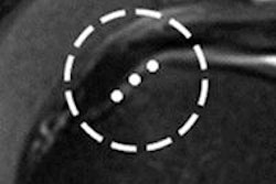
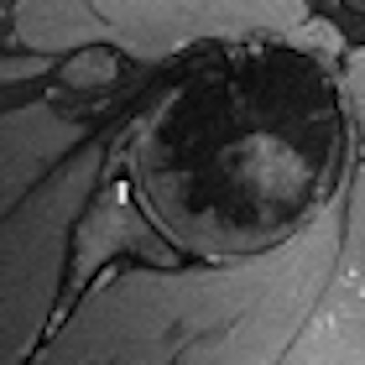
| MR (left) and MR arthrogram (right) are from the same patient with an anterior labral tear. The condition was repaired with surgical tacking. Images courtesy of Thomas Magee, MD. |
Arthroscopy detected 36 of the 38 posterior labral tears as fraying. Of those 36 patients, 19 were debrided, whereas 15 cases required no surgical intervention. Four cases of the 38 posterior labral tears had surgical tacking performed arthroscopically. Two cases were described as fraying by arthrogram.
Of the 28 anterior labral tears described on MR, 26 patients had surgical tacking performed. Two cases of the 28 had debridement.
Surgeons' perception
"The perception of the surgeons was that we were overcalling -- most specifically, SLAP tears," Magee concluded. "But it appears more of an issue of how we describe it, and what is the verbiage used in our reports and reports to the surgeons."
A high percentage of SLAP tears and posterior labral tears described in MR exams did not have surgical tacking performed, according to the researchers. Many of those cases were debrided with the primary purpose due to some other lesion. Most of the anterior labral tears had surgical tacking performed.
"Based on the results, our surgeons request we describe superior and posterior labral tears as fraying or tearing, unless we can clearly see a displaced tear," Magee said. "Most of the anterior labral lesions are treated surgically."
By Wayne Forrest
AuntMinnie.com staff writer
December 1, 2010
Related Reading
3T MRI as good as MR arthrography for planning hip surgery, February 1, 2010
Study: Rotator cuff treatment provides tendonitis relief, June 30, 2009
Noncontrast MRI effective in adhesive capsulitis diagnosis, June 3, 2009
Isotropic 3T MRI aids in diagnosing labrum, rotator cuff tears, December 29, 2008
MR arthrography outdoes 3-tesla MR on tendon tears, December 12, 2008
Copyright © 2010 AuntMinnie.com