Even if a negative CT scan shows no injuries in the cervical spine after blunt trauma, MRI should be used to evaluate patients who are obtunded or unexaminable to avoid missed injuries due to the modality's excellent sensitivity for soft-tissue injuries.
That conclusion comes from researchers at Harvard Medical School and Brigham and Women's Hospital in Boston, who found that CT, although highly sensitive in identifying abnormalities, cannot always find all clinically significant injuries.
MDCT "is incredibly sensitive and specific for picking up injuries, but the soft-tissue injuries that are not seen on CT scans can be catastrophic, even if they are of very low incidence," said co-author Dr. Mitchel Harris, chief of Brigham and Women's orthopedic trauma service. "MRI is exquisitely sensitive for all soft-tissue injuries -- ligaments, spinal cord, disk, and muscle injuries, all the things that cannot be readily identified on the CT scan."
Harris presented the findings at the recent American Academy of Orthopaedic Surgeons (AAOS) annual meeting. The lead author of the study is Dr. Andrew Schoenfeld, and the research also appeared in the January issue of the Journal of Trauma (Vol. 68:1, pp. 109-114).
Controversial issue
The authors wrote that ruling out injuries of the cervical spine in obtunded blunt trauma patients remains a "controversial issue."
"We have gotten very good at identifying bony injuries with high-level CT scans. We are still struggling with making sure we identify the nonbony injuries, the soft-tissue injuries," Harris said. "The natural inclination has been to order an MRI scan, but they are expensive. When trauma patients go to the MRI, they often are not medically stable. So, we are looking for other answers, other than throwing another test at them, to identify occult injuries."
The retrospective study analyzed several databases from 2000 to 2008, collecting results from 1,550 trauma patients with negative CT cervical spine scans who also were evaluated with MRI.
MRI detected a total of 194 abnormalities in 182 patients (12%). There were 86 ligament injuries (44%), and degenerative changes were identified in 47 cases (25%). Fractures and dislocations comprised the rest of the abnormalities found by MRI.
 |
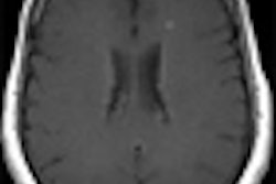
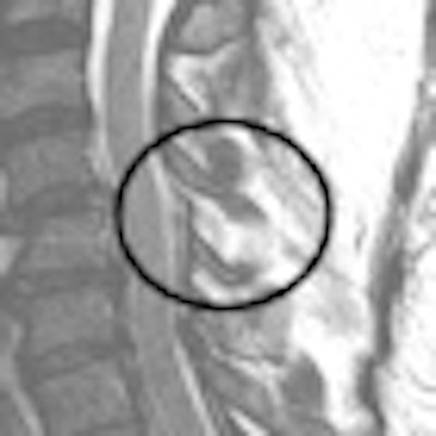
| Images of patient involved in a motor vehicle collision, who had head injury in addition to long-bone fractures of the lower legs. She was unable to participate in a reliable clinical exam. Her initial MDCT scan was negative for acute injury. |
 |
| MRI identified a posterior ligament injury, and the patient ultimately underwent surgery. All images courtesy of Dr. Mitchel Harris. |
Patient management
One of the more significant findings of the study is that the MRI results changed care management for 96 patients. "In 6% of cases, the MRI results altered patient care management, because the negative CT scan would have allowed the removal of the [cervical] collar," Harris said.
Among those 96 cases, 84 patients (5%) wore a cervical collar for an extended period of time, while 12 patients (1%) required surgery.
"One percent of the time in this study there was something on the MRI, even though the CT scan was negative, that made people decide to operate," Harris added. "Because it was a retrospective study and meta-analysis, I can't tell you what made those surgeons at that time decide that they needed an operation, but it matched the criteria" for the study.
Harris emphasized that CT is still the first modality of choice, as it is "clearly the best modality to pick up bony injuries, and the greater majority of injuries that occur are either bony or bony-plus. If you know you have a bony injury, you may be even more careful about treating the potential of an associated soft-tissue injury."
'Where it hurts'
A follow-up MRI is most beneficial for patients who cannot participate in a reliable CT exam due to multiple injuries, recent surgery, or a closed head injury, the study concluded. "Any situation where a patient can't tell you where it hurts," Harris said.
Harris and colleagues plan to expand their research to see if another imaging modality may replace MRI in certain trauma situations. "Is there another type of examination that we can do to avoid the MRI?" Harris said. "It's just not viable to keep spending $1,000 every time to determine [an injury]."
The researchers also would like to determine the true incidence of complete soft-tissue injuries of the spine. If physicians know beforehand that there is a less than 1% chance of a soft-tissue injury, Harris said, patients could be told there is a 99.5% chance there will be nothing adverse on the CT scan, and they can decide on their own how to proceed.
By Wayne Forrest
AuntMinnie.com staff writer
May 12, 2010
Related Reading
CT beats x-ray for pediatric cervical spine trauma, February 4, 2010
CT worth the radiation dose for traumatic C-spine injuries, October 30, 2009
Incidental findings common after cervical spine CT for trauma evaluation, March 17, 2009
CT more sensitive than spinal fracture x-ray, but has limits, January 30, 2006
CT: An all-star modality for C-spine trauma? August 18, 2004
Copyright © 2010 AuntMinnie.com