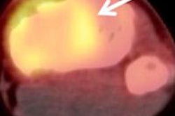
VIENNA - Diffusion-weighted MRI (DWI-MRI) stacks up well against PET/CT in a variety of oncology applications, such as tumor staging and the assessment of metastatic disease, according to presentations on the opening day of the European Congress of Radiology (ECR).
PET/CT has been the gold standard for many oncology applications, but whole-body diffusion-weighted MRI is beginning to show promise as a possible alternative modality. In a series of Thursday presentations, European researchers compared DWI-MRI to PET/CT, finding that while the modality performs well versus PET/CT, it's still not clear when and where each tool should be used.
In the first presentation, Dr. Carmelo Ciccio from the University of Rome "Tor Vergata" compared 3-tesla whole-body DWI-MRI to PET/CT for staging different types of malignant diseases. The group examined 60 patients with five different types of cancer between October 2008 and September 2009. The patient population had mean age of 63 years and a gender split of 39 men and 21 women.
The researchers compared DWI-MRI to three types of PET/CT techniques for N and M tumor staging: CT alone, PET and CT images viewed side-by-side, and fused PET/CT data. There was no statistically significant difference between DWI-MRI, which correctly staged 86% of patients, and fused PET/CT, which correctly staged 90%. The other techniques performed less well, with separate PET and CT correctly staging 70% of patients and CT alone 63%.
In the next presentation, Dr. Michael Fischer of University Hospital Zurich in Switzerland examined the utility of a software-based technique that fused DWI-MRI with CT, to match the MRI technique's functional information with the anatomical detail of CT.
The group was interested in substituting DWI-MRI for PET due to the latter's poor spatial resolution and anatomic information, but questions remained as to how suitable the technique was in a clinical environment. To answer these questions, the researchers compared fused DWI-MRI/CT with PET/CT in 52 patients with different types of malignant disease, both to assess the technique's performance and to see if it was feasible in a clinical setting.
Images were fused using a prototype software application in a two-step protocol. In the first step, DWI-MRI images with a b-value of 700 s/mm2 were displayed in an inverted grayscale. In the second step, diffusion-weighted images were semiautomatically fused with whole-body CT derived from a PET/CT study, yielding fused imaging. A team of readers assessed the images for precision of image fusion on a three-point scale, as well as diagnostic quality compared to PET/CT, also on a three-point scale.
The readers found the DWI-MRI/CT images to be poorly fused in 12 patients (23%), satisfactorily fused in 36 patients (69%), and correctly fused in four patients (8%). None of the readers rated DWI-MRI/CT images to be worse than PET/CT and nondiagnostic in any of the patients, while in 44 patients (85%) the images were rated to be worse than PET/CT but of diagnostic quality, and were rated to be equal to PET/CT in eight patients (15%).
Another presentation found that whole-body MRI could be used in the staging of lymphoma, and could be a better tool than PET/CT. A research team led by Dr. Goran Abdulgadhr from the University of Uppsala in Sweden examined 24 patients, comparing both modalities.
DWI-MRI and PET/CT performed the same in staging 20 of the patients, while different staging was the case in four patients. Of these four, three patients had indolent lymphoma (chronic lymphocytic leukemia/small lymphocytic lymphoma, or CLL/SLL), Abdulgadhr noted.
In a second presentation, the University of Zurich's Fischer returned to the podium to discuss a study in which DWI-MRI was used for detecting malignant tumors in 50 patients. The researchers used four different MRI techniques, including whole-body DWI, whole-body T2-weighted imaging, whole-body DWI with side-by-side T2 imaging, and whole-body DWI fused with T2 scans.
PET/CT was used as the reference standard for all studies. The performance of each technique was as follows:
|
As noted in the figures, combining T2 MRI with DWI-MRI significantly improved the diagnostic accuracy and detection rate of MRI, although there was no statistically significant difference whether the techniques were used in side-by-side fashion or as fused images, Fischer said.
Session moderator Dr. Fabian Kiessling of RWTH Technical University in Aachen, Germany, noted that the presentations highlighted the fact that DWI-MRI is a promising tool that shows equivalence to PET/CT for many applications -- the challenge is deciding when to use it.
"If one really wants to know which modality is better, we cannot take FDG-PET always as the gold standard," Kiessling said. "I think it might be important not only to compare these modalities, but in the future to look where they are complementary, to more focus on the points where we should use them together or use one or the other."
By Brian Casey
AuntMinnie.com staff writer
March 4, 2010
Related Reading
Pre-, postcontrast DWI-MRI similar in liver, spleen, and pancreas, May 18, 2009
MRI combination best at predicting progression after prostate cancer treatment, June 26, 2008
Diffusion-mapping MRI predicts radiation therapy response, April 22, 2008
Copyright © 2010 AuntMinnie.com