Texas researchers have published a case report demonstrating how medical imaging exams offer insight into the physiological changes wrought by the H1N1 virus in a 12-year-old girl who died of the disease.
Although the number of reported cases of H1N1 influenza is declining in most of the 209 countries it invaded, the World Health Organization (WHO) in Geneva reported on February 5, 2010, that H1N1 continues to be the predominant influenza virus circulating worldwide. The U.S. Centers for Disease Control and Prevention (CDC) estimates that between 830 and 1,730 children younger than 18 years of age died from symptoms related to the swine flu from April through December 2009.
At least one of these children developed acute necrotizing encephalopathy, according to a case report by radiologists at Driscoll Children's Hospital in Corpus Christi, TX, published in the February issue of Pediatric Radiology (2010, Vol. 40:2, pp. 200-205).
Because most pediatric fatalities are associated with pulmonary complications, pediatric radiologist Dr. Jane Lyon wanted to alert other radiologists to this particular case. Acute necrotizing encephalopathy is a rare condition, and most patients diagnosed with it have been Japanese children younger than 5 years who developed it as an unusual complication from influenza A virus.
The case described by Lyon and colleagues involved a 12-year-old girl who was admitted to Driscoll's emergency department on the day she developed the flu. Her symptoms upon hospital admission included a persistent fever as high as 104° F (40° C), profuse diarrhea, generalized pain, and increased weakness.
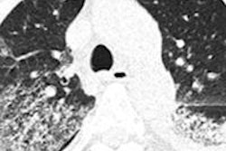
The patient had a normal chest x-ray that was performed when she was admitted to the hospital, as well as a normal-appearing CT exam. However, when an MRI exam was performed 14 hours later, it demonstrated multifocal signal abnormalities. Extensive symmetric restricted diffusion was identified in the thalami bilaterally. Restricted diffusion was also found in the periventricular white matter of the centrum semiovale, medial temporal lobes, and pontine gray nuclei, and symmetrically within the medial cerebellar hemispheres.
In addition, Lyon and colleagues identified an abnormally high T2 signal in areas of restricted diffusion, and more extensively in the brainstem and centrum semiovale.
 |
| Axial diffusion-weighted image (1000b), left, shows restricted diffusion in the thalami bilaterally. Axial T2-weighted image, right, demonstrates enlarged heterogeneous thalami with predominant T2 hyperintensity. All images courtesy of Driscoll Children's Hospital. |
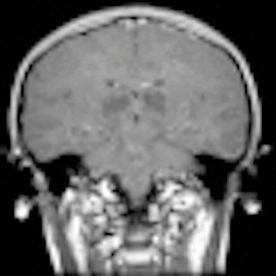
Abnormal rim enhancement of the bilateral thalami and magnetic susceptibility artifact in the bilateral thalami on T2-weighted images were also present. However, no abnormal meningeal enhancement was identified.
 |
| Contrast-enhanced coronal T1-weighted image, left, demonstrates abnormal enhancement in the centrum semiovale and brainstem with ring-enhancing lesions in the thalami bilaterally. Axial T2-weighted gradient recalled echo image, right, demonstrates magnetic susceptibility bilaterally in the thalami. |
A CT exam performed the next day showed abnormal bilaterally symmetric heterogenous attenuation in the thalami. Severe brainstem edema and progressive effacement of the basal cisterns developed, and on the third day of hospitalization, the patient had increased intracranial pressure with CT evidence of herniation and diabetes insipidus.
The patient was pronounced brain-dead. An autopsy did not identify any immunohistochemical or molecular evidence of influenza A in the brain. H1N1 influenza infection was confirmed in respiratory secretions, the authors reported.
By Cynthia E. Keen
AuntMinnie.com staff writer
February 12, 2010
Related Reading
Study categorizes x-rays of kids with swine flu, December 24, 2009
Chest CT depicts H1N1 better than radiography, October 21, 2009
CT reveals acute PE risk in patients with severe swine flu, October 21, 2009
CT can aid early swine flu diagnosis, October 14, 2009
Copyright © 2010 AuntMinnie.com