CHICAGO - High-pitch, low-contrast-dose dual-source CT angiography (CTA) can solve any number of challenges related to imaging patients who may need frequent follow-up scans of the abdomen and pelvis, according to a Tuesday presentation at RSNA 2016.
The technique could reduce key concerns surrounding contrast and radiation exposure for patients with renal impairment, especially.
The investigators from Northwestern University examined 20 patients referred for abdominopelvic CTA using a novel protocol that substantially reduced both radiation dose and contrast. Even with a dose as low as 3 mSv of radiation and 15 mL of iodinated contrast, the results showed no reduction in objective or subjective image quality using the technique.
"We showed that abdominal CT using an ultralow-dose contrast protocol is clinically and technically feasible, and we can achieve good image quality and homogeneous attenuation across vascular levels," said Dr. Faezeh Sodagari, who received an RSNA Student Travel Stipend Award for the CTA abstract.
Difficult-to-follow patients
CTA is frequently used to diagnose and follow-up various vascular diseases; however, the ability to administer contrast media is limited in patients with renal failure, or those at risk of contrast-induced nephropathy.
Newer generations of dual-source CT scanners offer wider detectors, lower tube potentials, and higher tube settings that can produce faster CTA studies at lower radiation doses, with far lower-than-standard contrast doses, Sodagari said.
"In our institution, we have developed a protocol which reduces the amount of contrast, and we wanted to know if an ultralow contrast media protocol ... has acceptable image quality and provides homogeneous attenuation across vascular levels in abdominal CT angiography," she said. "So we were very interested in this new study that recruited average-sized patients referred for abdominopelvic CT who received this new protocol, and we excluded patients with vascular thrombosis."
The study evaluated the homogeneity of vascular enhancement as well as image quality in 20 patients (mean age, 67 years) who underwent low-contrast-dose CTA at low kV.
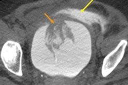
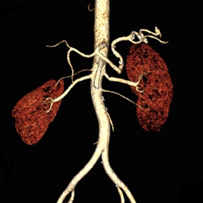
 Coronal (above, left), sagittal (above, right), and 3D reconstruction (below) images from abdominopelvic CTA in a 54-year-old woman who was scanned using 15 mL of iodinated contrast at 80 kV. Images courtesy of Dr. Faezeh Sodagari.
The acquisition protocol on a third-generation dual-source CT scanner (Somatom Force, Siemens Healthineers) included 0.6-mm collimation, 2.8 pitch (range, 1.6-3.2), tube current set by automatic exposure control, a scan duration of 0.8 seconds, and tube voltage of 80/90 kV with automatic tube voltage selection.
Before imaging, 15 mL to 30 mL of iodinated contrast (mean, 20.1 mL) diluted with saline was administered. Bolus tracking software triggered scan initiation at 130 HU in the abdominal aorta, and images were reconstructed using advanced modeled iterative reconstruction (ADMIRE, Siemens).
The investigators assessed intravascular contrast attenuation at the suprarenal aorta, infrarenal aorta, and right common iliac artery. They determined image noise, contrast-to-noise (C/N) ratio, and signal-to-noise (S/N) ratio. Sodagari and colleagues also extracted CT dose index volume (CTDIvol) from dose reports, and used repeated-measures analysis of variances for data analysis.
The mean CTDIvol was 3.8 ± 1.1, the mean C/N ratio was 9.2 ± 4.9, and the mean S/N ratio was 9.9 ± 4.1. Attenuation was homogeneous across vascular levels.
Regarding subjective image quality, 45% of the images were rated acceptable, 30% were good, and 25% were excellent, Sodagari said. Objective image quality parameters showed good C/N and S/N ratios, with relatively low noise levels considering the mean effective dose. Median CTDIvol was 3.4 at 80 kV and 3.6 at 90 kV.
| Attenuation, image noise, and C/N and S/N ratios | |||
| Suprarenal aorta | Infrarenal aorta | Right common iliac artery | |
| 80 kV (n = 16) | 217.7 ± 72.4 | 242.1 ± 55.8 | 244.4 ± 55.8 |
| 90 kV (n = 4) | 381.5 ± 121.1 | 380.2 ± 100.9 | 391.8 ± 103.8 |
| Image noise (HU) | 26.9 ± 9.2 | 27.9 ± 8.5 | 26.3 ± 7.7 |
| C/N ratio | 10.1 ± 5.8 | 10.9 ± 5.4 | 11.2 ± 5.8 |
| S/N ratio | 10.2 ± 5.3 | 10.5 ± 4.8 | 11.4 ± 5.7 |
Ultralow-dose contrast CTA with low radiation dose is both technically and clinically feasible, producing high image quality and homogeneous attenuation, Sodagari concluded.
The study shows the potential for reducing the contrast dose to as low as 15 mL for high-pitch abdominopelvic CT angiography at 80 kV. This may have clinical implications in abdominopelvic CT angiography, particularly in patients with renal impairment.