BOSTON - Certain non-small cell lung cancer (NSCLC) patients with clinical stage T2 tumors may still need their disease staged invasively, even if they have a negative PET/CT scan, according to a study presented on Sunday at the American Society for Radiation Oncology (ASTRO) meeting.
Researchers from Yale University found that the size of T2 NSCLC tumors, their location, and other characteristics could lead to evidence of stage 2 (N2) nodal metastases and may contradict a PET/CT indication of no nodal (N0) metastases. As a result, those patients may need to undergo invasive mediastinal staging or endobronchial ultrasound (EBUS) prior to curative therapy to confirm whether the disease has progressed.
"Centrally located tumors or solid T2 tumors should be strongly considered for further invasive staging, since the risk of occult mediastinal nodal involvement is greater than approximately 10%," said lead author Sarah Gao, a fourth-year medical student at Yale.
Diagnostic decisions
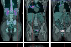
PET/CT traditionally has been the correct choice for staging nodal disease, which is considered the most important determinant of both management and prognosis, Gao told ASTRO attendees.
 Sarah Gao from Yale University.
"It also is becoming increasingly important now with the development of less invasive curative techniques, such as stereotactic body radiotherapy [SBRT] and wedge resection," she said. "The absence or presence of N2 nodes determines whether a patient is eligible for curative resection or SBRT. For all patients being considered for curative intensive therapy, PET/CT is recommended and thankfully has a sensitivity of 88% and specificity of up to 96% for nodal staging."
Based on staging results and the extent of disease, patients may undergo mediastinoscopy, which is the current gold standard for sampling upper and lower paratracheal nodes and metastases in the mediastinum. Another common alternative is endobronchial ultrasound, which is less invasive and may be better at reaching certain nodal locations.
Gao cited previous studies that found PET to be beneficial for nodal staging in NSCLC patients with clinical T1 and T2 tumors. In this patient population, PET's efficacy in finding occult nodal metastases in the mediastinal area ranges from 4% to 7%. Thus, National Comprehensive Cancer Network guidelines suggest that all patients with clinical stage I and II disease be staged by some form of invasive means. However, the criteria for surgical staging of these patients remain unclear, Gao said.
"This is a very broad heterogeneous group, and certainly there are some patients who are at greater risk for developing bone metastases than others," she said. "The question is: In this cohort of patients, are there certain patients with a greater risk for occult mediastinal nodal metastases than others, and who thus have a greater need for invasive staging? The goal of our study was to delineate a set of criteria that can help us say [which] patient is at risk for a nodal metastasis based on the staging, whereas another patient is at a lower risk and can therefore forego staging."
Retrospective analysis
The researchers retrospectively evaluated 329 consecutive patients who were diagnosed with clinical stage T1 to T2 tumors but had no evidence of nodal disease (N0). All subjects were being considered for curative therapy at Yale between 2011 and 2015. Of the total, 284 patients had their NSCLC staged by PET/CT, with 137 (48%) proceeding to mediastinoscopy, EBUS, or lobectomy followed by mediastinal dissection.
Following invasive NSCLC staging, 20 individuals (14%) were found to have occult N2 mediastinal nodal metastases. The negative predictive value for mediastinoscopy and EBUS was 96%, according to the researchers.
When they compared patients who developed N2 metastases with those who did not, they found significantly different characteristics related to tumor size, radiographic appearance, and tumor location. Based on tumor size, clinical T2 tumors had an 11.8% risk rate for N2 tumor metastases, with a risk rate of 3.8% for T1 tumors, Gao noted.
Centrally located tumors, which the Yale researchers defined as being within the inner two-thirds of the lung diameter, had an N2 metastases risk rate of 9.4%, compared with a rate of 1% for T1 tumors located on the periphery or outside the two-thirds of the lung diameter. Similarly, T2 tumors located within the inner one-third of the lung diameter had an N2 metastases risk rate of 17.5%, compared with 4.4% for T2 tumors located on the periphery beyond the inner one-third area.
Regarding radiographic appearance, solid clinical T2 tumors had a risk rate of 12.7% for progressing to N2 metastases, compared with a risk rate of 3.2% for T2 tumors with a ground-glass component.
"On the other hand, T1 tumors located on the outer one-third of the lung periphery and T2 tumors with any ground-glass component located on the outer one-third may not need staging because the rate of N2 tumor metastases is lower than 3%, and these patients may be able to safely forego mediastinoscopy and EBUS," Gao said.
The researchers also performed a univariate analysis that evaluated age, gender, maximum standardized uptake values, smoking history, and histology in relation to NSCLC. They concluded that none of those factors significantly contributed to the development of N2 metastases. The results were also confirmed through multivariate analyses.
The takeaway from the study is that NSCLC patients with centrally located or solid T2 tumors might have N2 nodal metastases, despite a negative or N0 finding on a PET/CT scan, Gao said.
"Regardless of whether we defined it as the inner one-third or inner two-thirds of the lung diameter, these patients had approximately 10% greater risk of mediastinal nodal metastases and should be considered for invasive staging," she said.