PET imaging of tau proteins with a novel radiotracer based on fluorine-18 (F-18) is showing great promise in differentiating between Alzheimer's patients and cognitively normal individuals and identifying how the disease may develop, according to a study published online July 25 in JAMA Neurology.
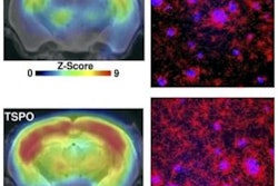
Researchers from Washington University in St. Louis found elevated levels of the tracer, F-18 AV-1451, in the hippocampus and cortical brain regions associated with Alzheimer's disease. The findings suggest that it takes a combination of tau deposits and beta-amyloid plaques for the neurodegenerative disease to progress.
"This work suggests that there is a progression of tau that starts in the lateral temporal lobe and spreads to additional areas," study co-author Dr. Beau Ances, PhD, wrote in an email to AuntMinnie.com. "We also demonstrated that F-18 AV-1451's standardized uptake value ratio in the hippocampus and Alzheimer's disease cortical signature regions can distinguish Alzheimer's from cognitively normal participants."
Alzheimer's contributors
As previous research has found, the initial accumulation of beta-amyloid plaques in the neocortical areas of the brain -- and its eventual spread to other regions -- often leads to Alzheimer's disease. The presence of tau proteins, which form with neurofibrillary tangles that first appear in the entorhinal cortex, can also contribute to the disease's progression.
 Dr. Beau Ances, PhD, from Washington University in St. Louis.
PET tracers have been developed to bind to beta amyloid and tau deposits to track the progression of the disease. One such imaging agent is AV-1451, which sticks to accumulated tau in the neurofibrillary tangles in neocortical brain regions of patients with Alzheimer's.
In fact, Ances et al in May published a paper that suggested tau protein deposits in the brain are a better indicator than beta-amyloid plaques of a person's progression to Alzheimer's disease. That study referred to the PET imaging agent T807, which is another name for AV-1451, Ances said.
The current study looks at the relationship between tau and cortical volumes, which are both components of neurodegeneration, according to Ances.
"This work tries to place tau imaging in relation to volumetric for staging of progression in sporadic Alzheimer's disease," he said.
AV-1451 targets
Ances and colleagues analyzed 38 men and 21 women with a mean age of 74 years (± 6 years) who were cognitively normal or who had Alzheimer's. Participants were eligible if they completed both AV-1451 PET imaging and brain MRI exams within 12 months of clinical assessment. The researchers excluded patients if they had neurologic, psychiatric, or systemic illness that might affect cognition, as well as if they had an autosomal-dominant mutation for Alzheimer's or other neurodegenerative disorders (JAMA Neurol, July 25, 2016).
Subjects underwent structural 3-tesla MRI scans (Magnetom Trio or Biograph mMR, Siemens Healthineers) and PET/CT scans (Biograph 40, Siemens). PET scans were performed 80 to 100 minutes after intravenous administration of approximately 9 mCi to 13 mCi of AV-1451.
Specifically, the researchers targeted AV-1451 binding in the hippocampus and Alzheimer's cortical signature regions with volumetric and cerebrospinal fluid (CSF) measurements to differentiate between Alzheimer's and cognitively normal participants. They also determined an AV-1451 standardized uptake value ratio (SUVR) of 1.19 within the cortical brain regions to separate beta-amyloid-positive Alzheimer's patients from beta-amyloid-negative cognitively normal subjects. The cutoff achieved sensitivity of 100% and specificity of 86%.
Based on the AV-1451 SUVR mark of 1.19 in the hippocampus and Alzheimer's-related cortical brain regions, the researchers were able to differentiate between patients with Alzheimer's disease and those who were cognitively normal. This same cutoff also distinguished between beta-amyloid-positive cognitively normal subjects with high and low levels of tau protein.
Beta-amyloid influence
Most interestingly, the presence of beta amyloid by itself was not related to hippocampal volume or the cortical thickness that is a signature of Alzheimer's disease. It took an elevated AV-1451 SUVR to indicate volumetric loss in both the hippocampus and Alzheimer's cortical signature regions in beta-amyloid-positive Alzheimer's patients versus cognitively normal participants who were positive or negative for beta amyloid.
"This suggests that a combination of both amyloid and tau are needed for progression in Alzheimer's," Ances said. "In particular, beta amyloid interacts with hippocampal and cortical tau pathology to affect neurodegeneration."
Using AV-1451 in staging the preclinical and clinical phases of Alzheimer's appeared to demonstrate that tau deposits in the hippocampus alone are not sufficient for the neurodegenerative process to lead to Alzheimer's.
"In particular, our results demonstrate that beta amyloid interacts with hippocampal and cortical tau pathology to affect neurodegeneration," Ances said. "It is the combination of both that leads to Alzheimer's."
Taking it one step further, beta amyloid "likely transforms pre-existing tauopathy to a more toxic species that results in neuronal injury," the authors wrote. "In the cerebral cortex, beta amyloid may intensify the spread of tauopathy, which in turn leads to neuronal loss that follows a topography that is similar to the observed spread of neurofibrillary tangles at autopsy."
Ances and colleagues plan to follow this research with longitudinal studies of tau using multiple biomarkers.