Software that fuses ultrasound images with MRI or CT studies can accurately guide liver biopsies for lesions that are hard to see on conventional ultrasound, yielding similar accuracy as CT-guided biopsy as well as some key benefits, according to an article in the June issue of the Journal of Ultrasound in Medicine.
In a retrospective study, a research team led by Dr. Yasmine Ahmed of University Hospitals Case Medical Center in Cleveland evaluated the performance of ultrasound fusion software, which enables interventional radiologists to view a patient's previously acquired CT or MR images side by side with the live ultrasound exam as they perform guided biopsies. Compared with CT-guided biopsy, ultrasound fusion-guided liver biopsy had a similar diagnostic yield, but it could be performed in about half the time and without exposing patients to additional radiation, according to Ahmed.
"Ultrasound fusion provides the same accuracy of CT[-guided biopsy], minus the ionizing radiation exposure," she told AuntMinnie.com.
Ultrasound's shortcomings
Ultrasound is the most cost-effective method for guiding various interventions in interventional radiology, but the modality has drawbacks such as low tissue contrast when compared with CT. That shortcoming can make it harder to locate and sample lesions, especially for less-experienced operators, Ahmed said.
 Dr. Yasmine Ahmed from University Hospitals Case Medical Center.
Because ultrasound fusion software (Smart Fusion, Toshiba America Medical Systems) was made available to their institution via a grant, the researchers decided to evaluate the software for guiding biopsy procedures of liver lesions that were poorly visible on ultrasound, she said. They compared these results with CT-guided liver biopsy procedures that had also been performed on lesions that were poorly visualized on ultrasound (J Ultrasound Med, June 2016, Vol. 35:6, pp. 1131-1141).
"We were aiming to find out whether the diagnostic accuracy and safety of ultrasound fusion was going to be comparable to, or higher than, that of CT-guided abdominal interventions," she said.
The researchers retrospectively reviewed 65 patients who were referred for a confirmatory liver biopsy following inconclusive diagnostic imaging of focal hepatic lesions between January 2012 and December 2014. The 65 subjects included all 35 patients who had received ultrasound fusion-guided liver biopsy in that time period, as well as a randomly selected sample of 30 patients who received CT-guided liver biopsy. Two patients in the CT group were later excluded, leaving 28.
All ultrasound fusion-guided biopsies were performed on an Aplio 500 scanner (Toshiba).
"When a limited screening evaluation of the liver using B-mode ultrasound showed lesions that were either invisible or hardly visible, a decision was made to switch to ultrasound fusion guidance instead," the authors wrote.
Matching anatomic landmarks on the real-time ultrasound feed with the fused CT/MR image took an additional five minutes for each procedure, according to the group.
CT-guided biopsies were performed on a Brilliance 3.6.2 big-bore CT system with a 16-slice configuration (Philips Healthcare). Unless otherwise contraindicated, patients received contrast-enhanced CT scans prior to the procedure to delineate the lesion for biopsy. The CT-guided biopsies were then performed with multiple sequential scans or via continuous observation using CT fluoroscopy, depending on the lesion's location and patient compliance.
"It is important to stress that, in our institution, choosing ultrasound fusion or CT to guide a biopsy of a focal hepatic lesion was reserved for cases that could not be performed under ultrasound guidance (e.g., obese patients and lesions that were hard to visualize with conventional ultrasound)," the authors wrote. "The choice to go for ultrasound fusion or CT relied on the expertise and preference of the available attending physician."
No yield difference
Ultrasound fusion-guided biopsy and CT-guided biopsy performed similarly in terms of diagnostic yield:
- Ultrasound fusion-guided biopsy: 32/35 (91.4%)
- CT-guided biopsy: 25/28 (89.3%)
For the purpose of the study, diagnostic yield was defined as procuring a histopathological sample that led to a conclusive diagnosis that aligned with follow-up imaging and did not require an additional biopsy, according to the researchers. The differences between the methods were not statistically significant (p = 0.88).
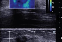
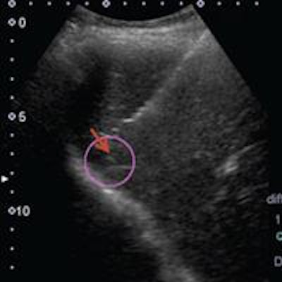
 Top: After a patient was referred for biopsy to further characterize a lesion with a nonspecific appearance on liver MRI, ultrasound fusion software was used to localize and biopsy the lesion. Bottom: The red arrow on the right panel points to the needle tip. All images courtesy of the Journal of Ultrasound in Medicine.
In other findings, ultrasound fusion-guided biopsy collected 101 core tissue samples from the 35 patients, compared with 54 core tissue samples in the 28 patients who received CT-guided biopsy. The difference was statistically significant (p < 0.0001).
The ultrasound fusion-guided biopsy also took much less time to perform:
- Mean procedure duration for ultrasound fusion-guided biopsy: 31.67 minutes ± 15.59 minutes
- Mean procedure duration for CT-guided biopsy: 61.67 minutes ± 26.54 minutes
That difference was also statistically significant (p = 0.003).
"Ultrasound fusion cuts the overall procedure time of image-guided liver biopsy [for] poorly visualized lesions on conventional B-mode ultrasound by approximately half the time when compared to CT," Ahmed said. "That would probably result in improved patient satisfaction and increase patient throughput."
In addition to requiring less time, ultrasound fusion-guided biopsy obviates the need to expose patients to radiation. Those who received CT-guided biopsy had an average radiation dose of 1,778.3 mGy-cm; the smallest dose was 386 mGy-cm and the largest was 4,131 mGy-cm.
The researchers also found that ultrasound fusion-guided biopsy had a mean largest lesion dimension of 2.91 cm. In contrast, the CT-guided biopsy group had a mean largest lesion dimension of 4.73 cm. The difference was statistically significant (p = 0.0001).
"These data suggest that the mean largest lesion dimension evaluated in the ultrasound fusion group was significantly smaller compared to the CT group," the authors wrote.
There were no significant differences in procedural complications. Two patients (5.7%) receiving ultrasound fusion-guided biopsy had pain sufficient to require analgesia, compared with one (3.6%) in the CT-guided biopsy group (p = 0.84).
"It's an overall safe procedure, with comparable accuracy to CT-guided liver biopsy," Ahmed said.
Usage advice
For those seeking to adopt ultrasound fusion software, Ahmed said it's important to always prepare and import the images that will be fused a day before the scheduled intervention. In addition, always fuse images that are less than 6 months old.
"Familiarize yourself with the software before your first intervention," she said. "[In addition], misregistration or misalignment can occur; thus, it is imperative that you familiarize yourself with the anatomy."