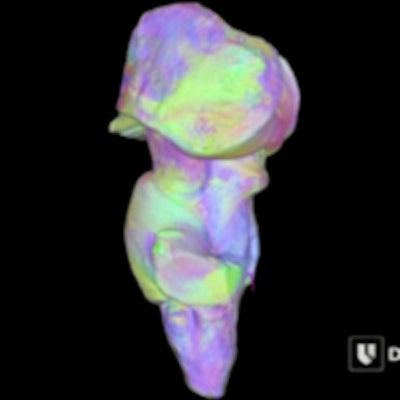
Researchers at Duke University have created an ultrahigh-resolution 3D MRI map of the human brainstem, intended to help surgeons use deep brain stimulation to treat patients.
The technology is expected to help surgeons implant electrodes in patients with conditions such as tremors and Parkinson's disease. Details of the research were published online June 3 in Human Brain Mapping.
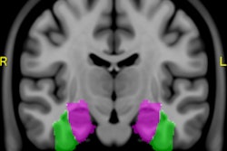
Study co-author Dr. Nandan Lad, PhD, director of the Duke NeuroOutcomes Center, said the 3D model greatly enhances the detail of brain regions such as the thalamus and the underlying circuitry being modulated.
 Scientists at Duke have produced a 3D map of the human brainstem. Image courtesy of Dr. Evan Calabrese, Duke Medicine.
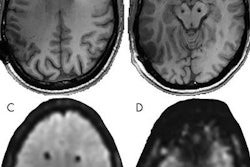
The map was derived from a 10-day scan of a healthy donor's postmortem brainstem using a 7-tesla MRI scanner. The images were converted into a 3D model that can be proportionally scaled to a patient's unique brain anatomy.
To assess the 3D map's accuracy, the researchers conducted a retrospective study of 12 patients who had been treated successfully for tremors using deep brain stimulation. The 3D model was used to determine the optimal placement of the electrodes in each patient. A predictive computer model and the actual successful electrode placements correlated for 22 of 24 electrodes in the dozen patients, the group found.
The researchers plan to begin a prospective study using the 3D model to guide deep brain stimulation surgery.