MRI at 3 tesla can help forecast the healing potential of a meniscus tear based, in part, on decreased meniscus extrusion, according to a study presented March 28 at a meeting of the American Orthopaedic Society for Sports Medicine (AOSSM).
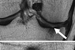
Researchers from the Rothman Institute in Philadelphia, led by Dr. Matthew Pepe, found that patients with recurrent meniscal root tears have an average meniscal extrusion of 1.5 mm, compared with an average meniscal extrusion of 1 mm in patients who healed properly.
The results indicate "significant stress on the area following the repair," the authors wrote. "Similar findings have also been shown in studies evaluating rotator cuff repair."
The meniscus provides stability in the leg, helps preserve the knee joint, and offers support for weight. The meniscus is also one of the most easily injured areas and is difficult to fully heal, the study authors noted.
Successful restoration of the meniscus is achieved primarily through healing in which the meniscus reattaches itself to the tibia. However, there is little information available on the healing of meniscus tears after a root repair, according to the researchers.
The study included five females and five males who had undergone a medial meniscus root repair by one surgeon who used an identical pullout surgical technique. The average follow-up time for scanning on a 3-tesla MRI system after surgery was 30 months, ranging from 21 to 41 months.
Pepe and colleagues also used the Western Ontario and McMaster Universities Osteoarthritis Index (WOMAC) and the Lysholm knee scoring scale to help quantify and compare outcomes. WOMAC and Lysholm help calculate the severity of pain, movement, function, and other mobility and comfort conditions of joints, such as knees and hips.
In this case, the measures were used to determine the quality of meniscus healing. Two fellowship-trained musculoskeletal radiologists then interpreted the MR images based on the study criteria.
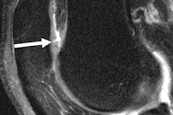
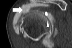
In the analysis, MR images found a new medial tear after the surgical repair in four patients. In addition, researchers found the recurrence of the meniscus root tear, or no healing of the root attachment, in four patients.
In the patients with a recurrent root tear, the meniscal extrusions averaged 1.5 mm, compared with average meniscal extrusions of 1 mm in patients who healed properly.
"There also was an increase in peripheral meniscus tears away from the repair in four patients, indicating excessive stress induced by the repair," the authors wrote.
Both WOMAC and Lysholm total scores range from 0 to 100, but higher scores with WOMAC denote worse conditions for patients, while higher scores with the Lysholm scale indicate better conditions. In the study, the average WOMAC score among all 10 subjects was 11.2 and the average Lysholm score was 81.6, but researchers found no correlation between the scores, meniscus healing, and clinical outcomes among the patients.
Pepe and colleagues concluded that successful repair and healing of meniscus were associated with decreased meniscus extrusion.
"However, this did not correlate with functional outcome scores, indicating that biologic healing is not a prerequisite for good clinical outcome," they wrote.
The additional knowledge on how meniscus tears heal will lead to improved clinical outcomes for all patients, they noted.