A common view is that chest x-rays are boring and difficult to read, but in fact spotting an abnormality on them brings huge satisfaction, and radiologists with excellent plain film skills often are highly regarded by colleagues, according to Irish researchers.
 Go forth and report plain films, urges Dr. Maria Twomey.
"Plain film interpretation is a key skill in becoming a competent independent radiologist and features heavily in examinations, yet it is often feared or neglected by radiology trainees," noted Dr. Maria Twomey and colleagues from the department of radiology at Cork and Mercy University Hospitals, Republic of Ireland, in an e-poster presented at RSNA 2014.
Tens of thousands of chest x-rays are performed every year in most hospitals and they are the "bread and butter" of radiology, yet they are seen by some people as not particularly relevant and interesting compared with MRI and CT. This is a serious mistake, they stated.
Their top general tips are as follows:
Practice by reporting plain films on a daily basis and taking responsibility as a resident/fellow to clear lists.
"Normal" is a powerful word that should be used it in your reports. Avoid cagey phrases like "no definite," which helps neither the referrer nor the patient. Don't be afraid to call a normal study normal, the authors emphasized.
Know your normal anatomy.
Old films are your best friend, and it's wrong to be too lazy to open them.
Understand the views and the rationale behind them.
Don't fear the lateral chest x-ray, because it's a great localizer.
Perform self-audit and peer review. For instance, randomly send every 20th radiograph to a colleague and vice-versa.
One view is too few.
Know "don't touch" lesions, such as nonossifying fibroma.
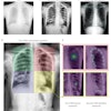
The crucial review areas are the lung apices, retrocardiac left lower lobe, and posterior recesses, the authors wrote. For the lateral chest x-ray, they think there are three golden rules: Vertebral bodies should get darker as you go more inferiorly, retrosternal space should be similar in density to the posteroinferior zone, and the heart should be relatively homogenous in density.
Interestingly, they presented an image that shows a deep brain stimulator and not a pacemaker with migrated or misplaced leads, as was reported.
Overall, their top "don't miss" items are listed as subtle right pneumoperitoneum, Rigler's sign (also known as the double wall sign), left upper lobe tumor on anteroposterior shoulder view, posterior fat-pad sign and effusion and supracondylar fracture in a pediatric elbow, C1 fracture and lateral dislocation, lateral mass C1 (overhanging C2), pneumomediastinum and subcutaneous emphysema, misplaced endotracheal tube and right upper lobe collapse, and left-sided Pancoast tumor.
Furthermore, in a hydropneumothorax, make sure you remember that pleural recesses are deeper than you think, and in the case of a deep sulcus sign, a pneumothorax on supine film can be very subtle, particularly in the emergency room, recommended Twomey et al.
"Plain films are interesting and challenging! Go forth and report!" they concluded.
Editor's note: The e-poster by Dr. Maria Twomey and colleagues received a Certificate of Merit award from the RSNA judges.